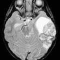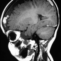
RADIOLOGY: HEAD: Case# 33617: SUBDURAL HEMATOMA SECONDARY TO CHILD ABUSE. This two year old child was brought to the emergency room with a history of "having just fallen down the stairs". T1 weighted coronal MRI reveals bilateral extra-axial fluid collections. Subdural hematomas are seen following significant craniocerebral trauma. They are especially common in cases of child abuse, and in elderly patients. They are thought to result from tearing of the cortical veins as they bridge the subdural space on their way to the dural sinus. The typical imaging appearance of an acute subdural hematoma is a crescent shaped extra-axial fluid collection seen diffusely over the hemisphere. They can usually be distinguished from epidural hematoma by this crescentic shape. Subdural hematomas also may be seen crossing skull sutures, while epidural hematomas do not. T2 wieghted axial MRI of a different patient reveals high signal chronic subdural hematomas bilaterally. Child abuse. T1 weighted sagital MRI of another patient shows mixed high and low signal of a subacute mixed with chronic subdural hematoma. Child abuse.
- Author
- Peter Anderson
- Posted on
- Thursday 1 August 2013
- Albums
- Visits
- 2034


0 comments