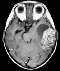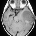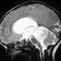
RADIOLOGY: HEAD: Case# 33616: PRIMARY CEREBRAL NEUROBLASTOMA (PRIMITIVE NEURO-ECTODERMAL TUMOR(PNET)). This 1 month old child presented with obtundation and a dilated pupil. T1 weighted axial MRI reveals a mixed signal mass in the left temporal lobe. T1 weighted axial MRI after gadolinium reveals enhancement of the lateral aspect, with a cystic component medially. T2 weighted axial MRI shows heterogeneous high signal in solid component of tumor. Primary cerebral neuroblastoma is considered one of the primitive neuroectodermal tumors (PNET). These tumors are a group of undifferentiated tumors arising in children with similar histologic features. These features include dense cellularity, with immature cells containing hyperchromatic nuclei and scant cytoplasm. There is an ongoing debate between the lumpers and splitters as to whether these tumors should be considered to represent a distinct group. Tumors considered to be a part of the PNET group (by the lumpers) include pineoblastoma, ependymoblastoma, medulloblastoma, medulloepithelioma, and primary cerebral neuroblastoma. Primary cerebral neuroblastomas are also referred to as supratentorial PNETs. They are tumors of infants and children, with most occurring before age five. Most are located in the frontal or parietal lobes, near the lateral ventricles, but they may occur anywhere within the CNS. Imaging studies reveal a large heterogeneous mass with variable enhancement. Cyst formation, hemorrhage, and calcification are common. CSF dissemination is frequently present, so the brain and spine should be screened for metastases with contrast enhanced MRI.
- Author
- Peter Anderson
- Posted on
- Thursday 1 August 2013
- Albums
- Visits
- 2060


0 comments