)
)
, occurring in about 10% of NF-I patients. This patient does have NF-I, but only about one fourth of patients with optic gliomas have NF-I. They commonly involve the optic chiasm and extend posteriorly to involve optic tracts and radiations, as in this case. Contrast enhancement is usually not as striking as in this case.")
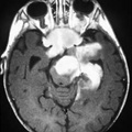
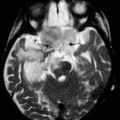
RADIOLOGY: HEAD: Case# 33615: OPTIC GLIOMA. This 4 year old child presented with decreased visiual acuity. T1 weighted axial MRI after gadolinium reveals an enhancing mass at the base of the brain extending into the orbital optic nerves, midbrain, and medial left temporal lobe. T1 weighted sagital MRI shows mass thickening the optic chiasm. T2 weighted axial MRI shows high signal mass. Optic gliomas are usually pilocytic astrocytomas. These tumors are one of the most common tumors in neurofibromatosis type I (NF-I), occurring in about 10% of NF-I patients. This patient does have NF-I, but only about one fourth of patients with optic gliomas have NF-I. They commonly involve the optic chiasm and extend posteriorly to involve optic tracts and radiations, as in this case. Contrast enhancement is usually not as striking as in this case.
- Author
- Peter Anderson
- Posted on
- Thursday 1 August 2013
- Tags
- head, neurofibromatosis, radiology
- Albums
- Visits
- 3094
0 comments