14/103
)
)

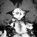
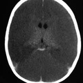
RADIOLOGY: HEAD: Case# 33612: EPIDURAL HEMATOMA. This four year old child was brought to the emergency room after he was hit by a car. CT scan without contrast obtained in the trauma center reveals a high density lens shaped collection in the left occipital region. Epidural hematomas are located between the dura and the skull. The tight adhesion of the dura to the skull causes the typical biconvex shape. This also accounts for the fact that epidural hematomas may cross dural attachments, but not skull sutures. Most commonly, they occur when a skull fracture lacerates the middle meningeal artery or a major dural sinus.
- Author
- Peter Anderson
- Posted on
- Thursday 1 August 2013
- Albums
- Visits
- 1732
0 comments