)
)

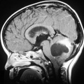
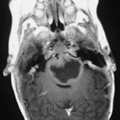
RADIOLOGY: HEAD: Case# 33609: CONTUSION SECONDARY TO CHILD ABUSE. This two year old child was brought to the emergency room with a history of "having just fallen of of her high chair". T1 weighted coronal MRI reveals an area of high signal within the posterior parietal lobe, with small bilateral extra-axial fluid collections. Cortical contusions occur with craniocerebral trauma when the brain strikes a bony ridge or dural fold. Most commonly this occurs in the tips and inferior surface of the temporal and frontal lobes, or in the parasagittal regions. MRI is frequently more sensitive in detecting these injuries. Subacute blood products in the contusion will appear bright on the T1 weighted image, as in this case. The presence of subdural hematoma and brain contusion should have raised your suspicion for child abuse.
- Author
- Peter Anderson
- Posted on
- Thursday 1 August 2013
- Albums
- Visits
- 1971
0 comments