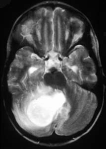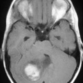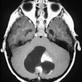
RADIOLOGY: HEAD: Case# 33608: CEREBELLAR PILOCYTIC ASTROCYTOMA. This two year old child presented with a history of ataxia and loss of motor coordination. T1 weighted axial MRI reveals a heterogeneous low signal mass in the right cerebellum. T1 weighted axial MRI after gadolinium shows enhancement of mass posterior to fourth ventricle. T2 weighted axial MRI shows high signal mass to be posterior to fourth ventricle. Pilocytic astrocytomas are slowly growing neoplasms of children and young adults which usually arise around the third and fourth ventricles. About one half arise in the optic pathways or hypothalamus, and one third arise in the cerebellar hemispheres or vermis. On imaging studies the cerebellar lesions are cystic with variably enhancing mural nodules.
- Author
- Peter Anderson
- Posted on
- Thursday 1 August 2013
- Albums
- Visits
- 1874


0 comments