)
)

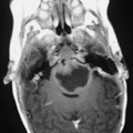
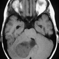
RADIOLOGY: HEAD: Case# 32947: BRAINSTEM GLIOMA. This 6 month old child presented to her family physician with a history of poor sucking and eye deviation. T1 weighted axial MRI after gadolinium reveals low signal mass expanding pons, with slight peripheral enhancement. T1 weighted sagital MRI shows mass within the pons, anterior to fourth ventricle. Brainstem gliomas are usually non pilocytic, low grade astrocytomas. On imaging studies they are most commonly solid and infiltrating, with variable contrast enhancement. Tumors intrinsic to the brainstem are biologically more aggressive than pilocytic astrocytomas arising in the optic pathways or cerebellar hemispheres, with most patients dying within two years. Occasionally cystic pilocytic astrocytomas may arise in this region.
- Author
- Peter Anderson
- Posted on
- Thursday 1 August 2013
- Albums
- Visits
- 2045
0 comments