2/7
)
)
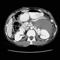
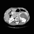
 Spontaneous left perinephric hematoma in this patient without history of trauma. Such a hematoma could occur secondary to a ruptured cyst or tumor. No tumor is definitely shown on this CT scan. Follow up CT scan is recommended after resolution of the hematoma in order to evaluate for a primary lesion in this area (three to six months). 2) Small left pleural effusion 3) Subcutaneous neurofibromas.")
RADIOLOGY: KIDNEY: Case# 33031: SPONTANEOUS PERINEPHRIC HEMATOMA & F/U EXAM. The patient is a 40-year-old black female with neurofibromatosis who presents with four days of back and abdominal pain.IVP showed what appeared to be a left renal pelvic filling defect. The patient denies any history of trauma. 1) Spontaneous left perinephric hematoma in this patient without history of trauma. Such a hematoma could occur secondary to a ruptured cyst or tumor. No tumor is definitely shown on this CT scan. Follow up CT scan is recommended after resolution of the hematoma in order to evaluate for a primary lesion in this area (three to six months). 2) Small left pleural effusion 3) Subcutaneous neurofibromas.
- Author
- Peter Anderson
- Posted on
- Thursday 1 August 2013
- Tags
- kidney, neurofibromatosis, radiology
- Albums
- Visits
- 2310
0 comments