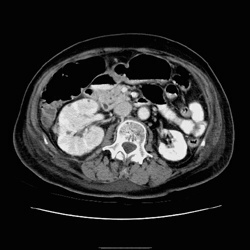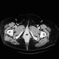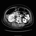
RADIOLOGY: KIDNEY: Case# 33016: PYELONEPHRITIS. 92 year old. 1. Large right kidney with findings consistent with pyelonephritis. 2. 1.2 cm right renal mass with enhancement pattern consistent with renal cell carcinoma. Suggest a repeat follow up CT scan after resolution of pyelonephritis to assure that this is a true finding and not in some way related to the pyelonephritis. There is a small soft tissue nodule adjacent to the proximal ureter, that probably represents an inflammatory lymph node, however would suggest follow-up of this at the same time to assure resolution. 3. Lytic area in the left posterior iliac wing, cannot rule out possible metastatic disease from renal cell carcinoma. Alternatively this may represent a small surgical defect from bone harvest, and clinical correlatin is suggested. 4. Mild central biliary dilatation of uncertain significance and suggest correlation with liver enzymes to rule out obstruction.
- Author
- Peter Anderson
- Posted on
- Thursday 1 August 2013
- Albums
- Visits
- 1342


0 comments