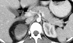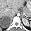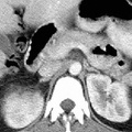
RADIOLOGY: ADRENAL: Case# 32907: F/U ADRENAL HEMMORHAGE. This is a 23 year old female who presentsfor follow-up of a large right adrenal mass, which was detected on MRI of the abdomen done seven months ago during her twenty-fifth week of gestation. The patient presented at that time with the sudden onset of right upper quadrant pain. She has been followed with ultrasound. The right adrenal mass has further decreased in size from 10.8 x 7.5 x 9.9 cm to 5 x 6 x 5.1 cm on todays exam. The mass is predominantly hypodense with minimally enhancing peripheral margin. A speck of high density is noted in the wall laterally which may represent an area of small calcification. Adrenal hemorrhage is most common in newborns usually following periods of hypoxia or trauma. In adults, adrenal hemorrhage usually results from trauma or infection and is usually unilateral. Bilateral hemorrhage is uncommon, but may occur following a bleeding diathesis, severe stress such as sepsis or major surgery or pregnancy, or during the first three weeks of anticoagulant therapy. Bilateral adrenal hemorrhage may result in a fatal adrenal insufficiency. Typical CT findings include bilateral hyperdense or soft tissue adrenal masses. When large, these may give the appearance of adrenal metastases;however, follow-up on CT will show reduction in size and density of an adrenal hemorrhage and calcifications as a late sequela. In addition, adrenal insufficiency is uncommon with metastatic disease. Post-traumatic hemorrhage involves the right gland 85% of the time. This may be explained by the elevation of the pressure in the inferior vena cava due to blunt trauma being more directly transmitted to the right gland as opposed to the left gland. CT typically show a hyperdense right adrenal mass with streaky infiltration of the periadrenal tissue, and enlargement of the right diaphragmatic crus.
- Author
- Peter Anderson
- Posted on
- Thursday 1 August 2013
- Tags
- adrenal, adrenal hemorrhage, radiology
- Albums
- Visits
- 2374


0 comments