)
)
. There is a 4.4cm transverse by 4.4cm AP lesion in the left lobe (image 22). A number of the lesions within the liver have central low attenuation. There is no adenopathy identified. There are no renal lesions. The bowel is displaced by the large liver, however, it appears unremarkable. The cervix is prominent, but otherwise unremarkable.")
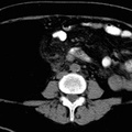
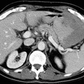
RADIOLOGY: HEPATOBILIARY: Case# 32890: MULTIPLE HEPATIC ADENOMAS. 32 year old woman with history of multiple masses within the liver. Biopsy at another institution revealed adenoma. This study is compared to a previous study. The lung bases are clear. There are multiple lesions within both the left and right lobe of the liver. A very small portion of the lateral segment of the left lobe appears spared. Overall, the liver measures 29cm cranio-caudal, with no appreciable change since the previous study. Representative focal lesions within the liver are unchanged in appearance or size. There is a 4.6cm transverse by 4.8cm AP lesion in the right lobe (image 14). There is a 4.4cm transverse by 4.4cm AP lesion in the left lobe (image 22). A number of the lesions within the liver have central low attenuation. There is no adenopathy identified. There are no renal lesions. The bowel is displaced by the large liver, however, it appears unremarkable. The cervix is prominent, but otherwise unremarkable.
- Author
- Peter Anderson
- Posted on
- Thursday 1 August 2013
- Tags
- hepatobiliary, radiology
- Albums
- Visits
- 767
0 comments