)
)
. She has known marked hepatosplenomegaly and has had prior splenic infarctions. There is moderately severe hepatomegaly without evidence of focal lesion or biliary dilatation. The spleen is massively enlarged measuring 25cm in cranio-caudal dimension. There is an irregular area of mixed predominantly low attenuation involving large wedge of tissue anteriorly and also extending upward toward the lateral upper splenic margin. Previously identified infarction in the lateral lower pole has resolved leaving a small scar. There is a trace of pelvic fluid. Chronic myeloid leukemia (CML), also referred to as chronic granulocytic leukemia (CGL), is a neoplastic clonal proliferation of myeloid stem cells. CML is characterized on a molecular level by a reciprocal chromosomal translocation between chromosomes 9 and 22, resulting in the formation of the Philadelphia chromosome. Other characteristics of CML include marked leukocytosis, with white cell counts varying from 50,000 to 200,000/?L; leukemic cells in the peripheral blood and bone marrow, mainly middle- to-late myeloid (granulocytic) precursor cells, including myelocytes, metamyelocytes, bands, and segmented forms; small numbers of blasts and promyelocytes; the Philadelphia chromosome, found in granulocytic and erythroid precursor cells and megakaryocytes; marked reduction in leukocyte alkaline phosphatase activity in the leukemic leukocyte. CML presents clinically with prominent splenomegaly and modestly enlarged liver and lymph nodes. The peak incidence of the disease is in the middle-age population (ages 35-50). In most cases the disease terminates in an accelerated phase known as a \"blastic crisis\" which is signaled by an increasing number of blast cells and promyelocytes. CT findings of CML include splenomegaly, hepatomegaly, and enlarged lymph nodes. Splenomegaly is defined as spleen size greater than 12 cm in the long axis and extension below the costal margin or substantially below the upper pole of the left kidney. Splenic infarction, a finding also asscoiated with CML, classically appers as a wedge-shaped hypodense area. However, the wedge shape is often obscured by the fusion of multiple infarcts of various sizes. The key finding is extension of the low density area to the capsule of the spleen without causing mass effect. The infarcted areas progressively atrophy, eventually resulting in notching of the splenic contour and occasionally in calcification. Other causes of splenic infarction include sickle cell and cardiac disease, lymphoma, and metastases. Splenic infarcts predispose patients for splenic abscess formation")
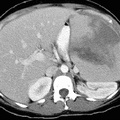
RADIOLOGY: ABDOMEN: Case# 32866: CML, SPLENIC INFARCT. Patient is a 30 year old female with chronic myeloid leukemia (CML). She has known marked hepatosplenomegaly and has had prior splenic infarctions. There is moderately severe hepatomegaly without evidence of focal lesion or biliary dilatation. The spleen is massively enlarged measuring 25cm in cranio-caudal dimension. There is an irregular area of mixed predominantly low attenuation involving large wedge of tissue anteriorly and also extending upward toward the lateral upper splenic margin. Previously identified infarction in the lateral lower pole has resolved leaving a small scar. There is a trace of pelvic fluid. Chronic myeloid leukemia (CML), also referred to as chronic granulocytic leukemia (CGL), is a neoplastic clonal proliferation of myeloid stem cells. CML is characterized on a molecular level by a reciprocal chromosomal translocation between chromosomes 9 and 22, resulting in the formation of the Philadelphia chromosome. Other characteristics of CML include marked leukocytosis, with white cell counts varying from 50,000 to 200,000/?L; leukemic cells in the peripheral blood and bone marrow, mainly middle- to-late myeloid (granulocytic) precursor cells, including myelocytes, metamyelocytes, bands, and segmented forms; small numbers of blasts and promyelocytes; the Philadelphia chromosome, found in granulocytic and erythroid precursor cells and megakaryocytes; marked reduction in leukocyte alkaline phosphatase activity in the leukemic leukocyte. CML presents clinically with prominent splenomegaly and modestly enlarged liver and lymph nodes. The peak incidence of the disease is in the middle-age population (ages 35-50). In most cases the disease terminates in an accelerated phase known as a "blastic crisis" which is signaled by an increasing number of blast cells and promyelocytes. CT findings of CML include splenomegaly, hepatomegaly, and enlarged lymph nodes. Splenomegaly is defined as spleen size greater than 12 cm in the long axis and extension below the costal margin or substantially below the upper pole of the left kidney. Splenic infarction, a finding also asscoiated with CML, classically appers as a wedge-shaped hypodense area. However, the wedge shape is often obscured by the fusion of multiple infarcts of various sizes. The key finding is extension of the low density area to the capsule of the spleen without causing mass effect. The infarcted areas progressively atrophy, eventually resulting in notching of the splenic contour and occasionally in calcification. Other causes of splenic infarction include sickle cell and cardiac disease, lymphoma, and metastases. Splenic infarcts predispose patients for splenic abscess formation
- Author
- Peter Anderson
- Posted on
- Thursday 1 August 2013
- Albums
- Visits
- 1136
0 comments