)
)

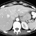
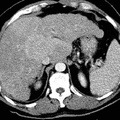
RADIOLOGY: HEPATOBILIARY: Case# 32864: CIRRHOSIS, HCC. A 54 year old white male with history of alcohol abuse with cirrhosis. Phase CT shows borderline hepatomegaly with the liver measuring 18 cm in length. The liver has a nodular contour suggesting cirrhosis. The liver demonstrates abnormal patchy heterogeneous contrast enhancement diffusely. There are multiple ill-defined areas of low attenuation areas throughout the liver. These are better seen on the delayed images. There is a 7 x 7 cm mass in the inferior aspect of the posterior segment of the right lobe of the liver. The main portal vein appears unremarkable without evidence of thrombus. The intrahepatic IVC is partially effaced. Multiple serpiginous tubular structures are identified in the region of the splenic hilum extending to the left kidney, representing varices. Although uncommon in the United States, hepatocellular carcinoma is an important cause of death in parts of Africa and Asia because of the hepatotrophic viruses. In the United States, eighty percent of hepatocellular carcinomas arise in cirrhotic livers. Three patterns of tumor growth are seen: 50% present as a solitary tumor, 30% as a diffuse infiltrative tumor, and 20% as a multinodular tumor. The tumor usually appears as a hypodense or isodense lesion on nonenhanced images and enhances prominently during the arterial phase on dynamic contrast injection. Areas of tumor necrosis and calcification are common. Tumor invasion of hepatic and portal veins occurs frequently.
- Author
- Peter Anderson
- Posted on
- Thursday 1 August 2013
- Tags
- hepatobiliary, radiology
- Albums
- Visits
- 746
0 comments