)
)

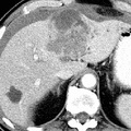
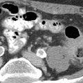
RADIOLOGY: HEPATOBILIARY: Case# 32855: HEP. MASS. This is a 74 year old male with acute onset of abdominal pain over 24 hours. Outside non-contrast CT scan demonstrated a left hepatic lobe lesion. We are requested to evaluate further with contrasted CT. There is a 6 x 7 cm heterogeneous predominantly low attenuation mass in the medial segment of the left hepatic lobe. The mass appears to rupture through the liver capsule anteriorly and results in a moderate amount of intraperitoneal hemorrhage. Some of the hemorrhage adjacent to the mass is of higher attenuation consistent with a sentinel clot. There is some biliary dilatation in the lateral segment of the left lobe and this segment of the liver appears somewhat atrophic. There are other more well defined low attenuation areas within the liver in the lateral segment of the left lobe and the posterior segment of the right lobe which are felt to represent cysts. There is a focal area of oral contrast, suspicious for a gastric ulcer, either benign or malignant in etiology.
- Author
- Peter Anderson
- Posted on
- Thursday 1 August 2013
- Tags
- hepatobiliary, radiology
- Albums
- Visits
- 805
0 comments