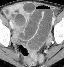
RADIOLOGY: GASTROINTESTINAL: GI: Case# 32851: INCARCERATED INGUINAL HERNIA. This is a 62 year old female status post partial gastrectomy and gastroenterostomy (approximately 35 years ago for duodenal ulcer disease), and status post hysterectomy and left inguinal herniorrhaphy, presents with nausea and vomiting and mild leukocytosis. CT is done to evaluate for abscess. No prior abdominal CTs for comparison. There are multiple loops of dilated small bowel which extend inferiorly to a region in the lower right anterior abdominal wall with a focal loop of incarcerated small bowel, which is the transition point. A few loops of small bowel more distally are decompressed and the colon is not dilated. This appearance is consistent with a distal high grade ileal small bowel obstruction due to an incarcerated loop of small bowel in a right inguinal hernia. Status post hysterectomy. There is mild increased density throughout the mesentery consistent with mild mesenteric edema. The IVC is flattened in its mid abdominal course likely due to the pressure from the enlarged loops of small bowel. The duodenum (afferent loop) is decompressed.