)
)
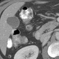
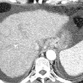
. There is a retrocecal appendix which courses cephalad. The tip of the appendix lies in the hepatorenal fossa, and contains barium. No inflammatory change of the surrounding fat is noted. The appendix is not always located directly under McBurneys point. It may be anterior or posterior; it may be long or short; it may be mobile or relatively fixed in position. Retrocecal locations as shown above are not uncommon. At UAB, we have even seen an appendix located in an inguinal hernia!")
RADIOLOGY: GASTROINTESTINAL: GI: Case# 32842: RETROCECAL/RETROHEP. APPENDIX. This is a symptomatic 57 year old black male with a small, indeterminate right hepatic lobe lesion by recent ultrasound. The cecum is relatively cephalad in location, located just below the inferior margin of the liver (either secondary to a mobile cecum or incomplete rotation). There is a retrocecal appendix which courses cephalad. The tip of the appendix lies in the hepatorenal fossa, and contains barium. No inflammatory change of the surrounding fat is noted. The appendix is not always located directly under McBurneys point. It may be anterior or posterior; it may be long or short; it may be mobile or relatively fixed in position. Retrocecal locations as shown above are not uncommon. At UAB, we have even seen an appendix located in an inguinal hernia!
- Author
- Peter Anderson
- Posted on
- Thursday 1 August 2013
- Tags
- gastrointestinal, radiology
- Albums
- Visits
- 765
0 comments