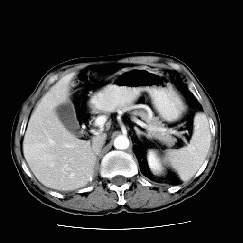
RADIOLOGY: HEPATOBILIARY: Case# 32822: RCC METASTASIS, HEMANGIOMA. This is a 62 year old male who is three years status post right radical nephrectomy for a renal cell carcinoma. The patient had a previous abdominal CT done two months ago that showed a suspicious liver mass. Pre-contrast images through the liver demonstrate a 2 x 1.5cm low attenuation mass inferiorly and posteriorly which was identified on the previous CT and a 4 x 4cm low attenuation mass within the posterior segment of the right lobe not previously identified. The 4cm mass shows intense but fairly homogenous peripheral enhancement with central low attenuation. Delayed images through the liver show central filling of the large mass. The smaller mass is not visible on post contrast images. The appearance of the small lesion is grossly unchanged from the prior study when it was thought to be a hemangioma. The liver is a common site for the metastatic spread of malignant tumors. Metatstatic lesions are the most common malignant tumor of the liver. Most arise from the colon, although they may originate from almost any primary malignancy. Liver metastases are usually multiple, well-defined, low density solid masses. When there is fatty infiltration, the masses may appear as high density solid masses. When the tumor is mucinous colon or ovarian carcinoma, melanoma, lung, or carcinoid, the tumor may have a cystic or necrotic appearance. Metastatic lesions may also be calcified. A solitary metastatic lesion may present a problem in differentiation. Thus it is necessary to consider the other possibilities such as hepatoma, hepatic adenoma, focal nodular hyperplasia, and cavernous hemangioma. Cavernous hemangiomas are the second most common focal mass found in the liver. They are usually asyptomatic, however when very large, may cause symptoms by pressure effect, hemorrhage, or arteriovenous shunting. The classic CT findings include a well-defined hypodense mass of same density of other blood-filled spaces, the development of nodule-like areas of enhancement from the periphery following administration of a bolus of contrast, areas of enhancement become confluent and isodense of hyperdense relative to the liver parenchyma, prolonged retention of contrast material, persisting within the lesion for 20 to 30 minutes, and discrete areas of fibrosis and calcification which remain hypodense throughtout the enhancement period.