)
)
 include pelvic muscles, bladder, prostate, seminal vesicles, and ovaries.")
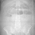
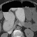
RADIOLOGY: GASTROINTESTINAL: GI: Case# 32820: ADENO. OF THE COLON. 46 year old female status post hysterectomy and bilateral salpingo-oophrectomy two weeks prior. She now presents with abdominal pain, nausea, and vomiting. There is a 4 x 4 cm soft tissue mass with a central hypodensity in the region of the splenic flexure of the colon. The colon proximal to the mass is dilated and fluid-filled. The distal colon is collapsed. Adenocarcinoma of the colon appears as an irregularly marginated, roughly spherical soft tissue mass, as seen in the transverse colon above. Larger tumors may demonstrate central low attenuation representing necrosis. Lesions of the rectum and rectosigmoid are seen as asymmetric or circumferential thickening of the bowel wall with deformation and narrowing of the lumen. Other findings include: extension of the tumor intopericolonic fat, invasion of adjacent structures, lymphadenopathy, adrenal or liver metastases, hydronephrosis, ascites, and masses in the abdominal wall, omentum or mesentery. Common sites of local extension from the rectosigmoid (as seen by marginal obscuration) include pelvic muscles, bladder, prostate, seminal vesicles, and ovaries.
- Author
- Peter Anderson
- Posted on
- Thursday 1 August 2013
- Tags
- gastrointestinal, radiology
- Albums
- Visits
- 887
0 comments