
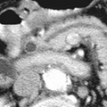
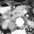
RADIOLOGY: HEPATOBILIARY: Case# 32810: COMMON BILE DUCT STONES. 67 year old male with a three month history of weight loss and abdominal pain. Simple appearing hepatic cysts are present, one in the left lobe measuring 2.1cm and two in the inferior portion of the right lobe, the largest 3.1cm in size. There is mild intrahepatic biliary ductal dilatation. The common duct is dilated to 8mm and contains multiple concentric high attenuation stones near the ampulla. Multiple stones are present in the gallbladder. The pancreatic duct is mildly dilated to 3mm. No mass is seen in the pancreas. The right adrenal gland contains a 1.6 x 1.6cm low attenuation mass. The spleen is 14cm in cephalocaudad length without focal lesions. The aorta is heavily calcified with mural thrombus and a maximum AP diameter of 3.1cm. Although common bile duct stones may form de novo in the biliary tree, most come from the gallbladder. CT is about 95% accurate in diagnosing biliary obstruction. CT diagnosis is based on the findings of dilated intra- and extrahepatic bile ducts. This dilatation may be absent in cirrhosis, cholangitis, or periductal fibrosis, thereby creating a false-negative. Dilated intrahepatic ducts will appear as branching tubular or round structures of low density enlarging as they approach the porta hepatis. Dilatation of the common bile duct to greater than 7mm in diameter also indicates obstruction. CT is also useful in differentiating causes of obstruction. Abrupt tapering of a dilated duct suggests a malignant process, such as pancreatic carcinoma or cholangiocarcinoma, whereas gradual tapering is more commonly seen with an inflammatory process such as chronic pancreatitis. Gallstones are visualized as calcified or soft tissue density structures within the bile duct surrounded by lower density bile creating a target sign. If the stone is in the dependent part of the bile duct, this will create a crescent-shaped area of bile anteriorly. Some stones are isodense with bile and therefore not detectable by CT.
- Author
- Peter Anderson
- Posted on
- Thursday 1 August 2013
- Tags
- hepatobiliary, radiology
- Albums
- Visits
- 839
0 comments