)
)

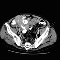
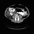
RADIOLOGY: GASTROINTESTINAL: GI: Case# 117: APPENDICITIS. This sixty year old woman has severe multiple sclerosis and presents with acute abdominal pain. There is an approximately 2 x 1 cm metallic density with a somewhat irregular contour noted in the region of the cecum, probably within the lumen of the bowel. There is extensive inflammation in the right lower quadrant with hazy changes and adherent small bowel loops with thickened bowel wall and mild distention of the bowel in this region. Free fluid is identified in the right paracolic gutter and pelvis. No free air is identified, nor is there evidence of a bowel obstruction. On image #37, a very thickwalled appendix is noted, with a particularly bulbous appendiceal tip accompanied by hazy inflammatory changes.
- Author
- Peter Anderson
- Posted on
- Thursday 1 August 2013
- Tags
- gastrointestinal, radiology
- Albums
- Visits
- 1009
0 comments