)
)

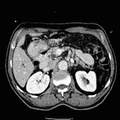
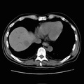
RADIOLOGY: HEPATOBILIARY: Case# 113: COMMON BILE DUCT STONE @ AMPULLA. 68 year old male with painless jaundice. 1. Intrahepatic ductal dilatation with dilated common bile duct to the area of the duodenum and in this area a mass is noted. This mass likely represents either a periampullary pancreatic mass or a cholangiocarcinoma. ERCP would be helpful to further evaluate this lesion. No evidence of choledocholithiasis. 2. Area of low attenuation within the left common femoral vein was seen. The patient was then taken to ultrasound. After further investigation, there was no evidence of a deep venous thrombosis. This area of low attenuation was most likely due to volume averaging. 3. Status post cholecystectomy. 4. No evidence of focal liver lesions or osseous metastases.
- Author
- Peter Anderson
- Posted on
- Thursday 1 August 2013
- Tags
- hepatobiliary, radiology
- Albums
- Visits
- 992
0 comments