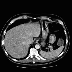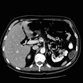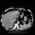
RADIOLOGY: ABDOMEN: Case# 110: GLUCAGONOMA -POSITIVE OCTREOTIDE. The patient is a 60 year old white male with a peculiar rash and diarrhea. A 2 x 2.5 mildly heterogeneous mass is seen at the splenic hilum in the pancreatic tail. Otherwise, the pancreas is unremarkable. A 2.5 x 1.5 cm low attenuation lesion is seen in the liver adjacent to the falciform ligament. A second low attenuation measuring less than 1 cm is seen in the medial segment of the left hepatic lobe and is indeterminate due to size. The spleen is normal in appearance without focal lesions present. A small approximately 1 cm accessory spleen is seen lateral to the splenic hilum. Glucagonoma is one of the pancreatic islet cell tumors. The rash associated with glucagonoma is necrolytic migratory erythema.
- Author
- Peter Anderson
- Posted on
- Thursday 1 August 2013
- Albums
- Visits
- 1777


0 comments