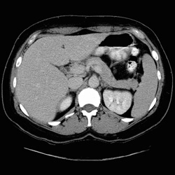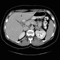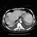
RADIOLOGY: PANCREAS: Case# 108: PANCREATIC HEAD INSULINOMA. 42-year-old white female with hypoglycemia. A vascular lesion was seen angiographically in the pancreatic neck. There is a slight prominence of the portal confluence and prominent common bile duct which tapers smoothly. Other than for a small anterior protuberance of a portion of the parenchyma within the pancreatic neck, no pancreatic abnormalities are seen. Specifically, the enhancing mass demonstrated angiographically is not discernable. Islet cell tumors, such as insulinoma, present much sooner than their adenocarcinoma counterparts due to the symptom-causing secreted hormones. As a consequence, islet cell tumors are generally very small at presentation with most less than 2 cm in size. Islet cell tumors are hypervascular; therefore, they enhance briskly on contrast CT and may be demonstrated with angiography. CT can detect approximately 75% of islet cell tumors, but only about 50% of insulinomas.
- Author
- Peter Anderson
- Posted on
- Thursday 1 August 2013
- Albums
- Visits
- 1176


0 comments