)
)
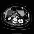
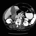
: 82 year old female with abdominal pain and palpable abdominal mass. Exam 1: Inflammatory changes along the ascending colon from the level of the appendix to the hepatic flexure. In addition, there is some mild thickening of the ascending colonic wall. Free intraperitoneal air is present. Exam 2: There is a large complex mass present in the right abdomen extending from the inferior aspect of the liver into the pelvis. Inflammatory changes involve most of the adjacent structures within the abdomen. The hallmark of colonic perforation on CT is focal pericolonic inflammation, as evidenced by diffuse or streaky opacities in the pericolonic fat due to edema, hyperemia, and cellular inflammation. Adenocarcinoma of the colon appears as an irregularly marginated, roughly spherical soft tissue mass, as seen in the right colon above. Larger tumors may demonstrate central low attenuation representing necrosis. Other findings include: extension of the tumor intopericolonic fat, invasion of adjacent structures, lymphadenopathy, adrenal or liver metastases, hydronephrosis, ascites, and masses in the abdominal wall, omentum or mesentery.")
RADIOLOGY: GASTROINTESTINAL: GI: Case# 98: PERFORATED COLON CA WITH ABSCESS. Exam 1: 82 year old female with abdominal pain. Exam 2 (6 mos.later): 82 year old female with abdominal pain and palpable abdominal mass. Exam 1: Inflammatory changes along the ascending colon from the level of the appendix to the hepatic flexure. In addition, there is some mild thickening of the ascending colonic wall. Free intraperitoneal air is present. Exam 2: There is a large complex mass present in the right abdomen extending from the inferior aspect of the liver into the pelvis. Inflammatory changes involve most of the adjacent structures within the abdomen. The hallmark of colonic perforation on CT is focal pericolonic inflammation, as evidenced by diffuse or streaky opacities in the pericolonic fat due to edema, hyperemia, and cellular inflammation. Adenocarcinoma of the colon appears as an irregularly marginated, roughly spherical soft tissue mass, as seen in the right colon above. Larger tumors may demonstrate central low attenuation representing necrosis. Other findings include: extension of the tumor intopericolonic fat, invasion of adjacent structures, lymphadenopathy, adrenal or liver metastases, hydronephrosis, ascites, and masses in the abdominal wall, omentum or mesentery.
- Author
- Peter Anderson
- Posted on
- Thursday 1 August 2013
- Tags
- gastrointestinal, radiology
- Albums
- Visits
- 1252
0 comments