)
)
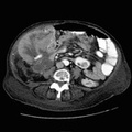
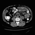
 and loss of the normal rugal folds may be seen on CT imaging. The abnormally thickened wall may exhibit enhancement after contrast administration. Gastric carcinoma spreads via four mechanisms: [1] hematogenous dissemination to liver and lungs [2] direct extension to porta hepatis (along gastrohepatic ligament) and transverse colon (along gastrocolic ligament) [3] lymphatic spread to regional nodes [4] intraperitoneal spread to peritoneum and peritoneal surfaces of bowel.")
RADIOLOGY: GASTROINTESTINAL: GI: Case# 95: GASTRIC ADENOCARCINOMA. 68 year old male with epigastric pain. There is a large, fungating soft tissue mass protruding off of the lesser curvature. The gastric cardia and proximal fundus are narrowed by the mass. Gastric adenocarcinoma is the third most common GI malignant neoplasm. Most gastric cancers arise in the distal stomach. Morphologic types include exophytic, diffusely infiltrative, and ulcerating. Wall thickening (with an adequately distended gastric lumen) and loss of the normal rugal folds may be seen on CT imaging. The abnormally thickened wall may exhibit enhancement after contrast administration. Gastric carcinoma spreads via four mechanisms: [1] hematogenous dissemination to liver and lungs [2] direct extension to porta hepatis (along gastrohepatic ligament) and transverse colon (along gastrocolic ligament) [3] lymphatic spread to regional nodes [4] intraperitoneal spread to peritoneum and peritoneal surfaces of bowel.
- Author
- Peter Anderson
- Posted on
- Thursday 1 August 2013
- Tags
- gastrointestinal, radiology
- Albums
- Visits
- 1179
0 comments