)
)
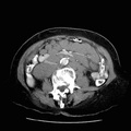
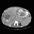
. Metastases may become necrotic with low attenuation centers and/or display calcifications (which may become extensive).")
RADIOLOGY: ABDOMEN: Case# 91: COLON CANCER, CALCIFIED. A 43 year old female with a history of colon carcinoma with metastatic disease diagnosed 2 months ago. She is status post hepatic artery chemotherapy with FUDR two weeks ago. Multiple hypodense lesions with extensive calcifications are seen throughout all segments of the liver. There is gross hepatomegaly. The enlarged liver compresses the stomach and displaces the pancreas and duodenum laterally. Splenic varices and splenomegaly are present. Hepatic metastases vary in their CT apperance. Borders may be sharp, ill-defined, or nodular with round, ovoid, or irregular shape. Attenuation is usually lower than that of the surrounding liver both before and after contrast administration (although mets may become iso- or hyperdense after contrast). Metastases may become necrotic with low attenuation centers and/or display calcifications (which may become extensive).
- Author
- Peter Anderson
- Posted on
- Thursday 1 August 2013
- Albums
- Visits
- 1495
0 comments