)
)
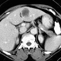
 include pelvic muscles, bladder, prostate, seminal vesicles, and ovaries. Hepatic metastases vary in their CT apperance. Borders may be sharp, ill-defined, or nodular with round, ovoid, or irregular shape. Attenuation is usually lower than that of the surrounding liver both before and after contrast administration (although mets may become iso- or hyperdense after contrast). Metastases may become necrotic with low attenuation centers and/or display calcifications.")
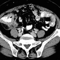
RADIOLOGY: GASTROINTESTINAL: GI: Case# 83: COLON CA, WITH HEPATIC METS. Patient is a 70 year old female with colon carcinoma diagnosed one month ago by colonoscopy for pre-operative staging. There is a 3.5x3.0cm low attenuation lesion in the lateral segment of the left lobe of the liver. There is no evidence of biliary dilatation. Spleen, adrenals, kidneys, pancreas, and stomach and small bowel are normal. There is irregular thickening of the cecum and right colon with a few minimal strandy changes in the adjacent fat. The adjacent appendix is also dilated and fluid-filled. No adenopathy is identified. Adenocarcinoma of the colon appears as an irregularly marginated, roughly spherical soft tissue mass, as seen in the right colon above. Larger tumors may demonstrate central low attenuation representing necrosis. Lesions of the rectum and rectosigmoid are seen as asymmetric or circumferential thickening of the bowel wall with deformation and narrowing of the lumen. Other findings include: extension of the tumor intopericolonic fat, invasion of adjacent structures, lymphadenopathy, adrenal or liver metastases, hydronephrosis, ascites, and masses in the abdominal wall, omentum or mesentery. Common sites of local extension from the rectosigmoid (as seen by marginal obscuration) include pelvic muscles, bladder, prostate, seminal vesicles, and ovaries. Hepatic metastases vary in their CT apperance. Borders may be sharp, ill-defined, or nodular with round, ovoid, or irregular shape. Attenuation is usually lower than that of the surrounding liver both before and after contrast administration (although mets may become iso- or hyperdense after contrast). Metastases may become necrotic with low attenuation centers and/or display calcifications.
- Author
- Peter Anderson
- Posted on
- Thursday 1 August 2013
- Tags
- gastrointestinal, radiology
- Albums
- Visits
- 1534
0 comments