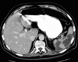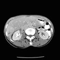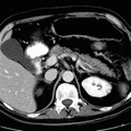
RADIOLOGY: SPLEEN: Case# 82: SPLENIC & RENAL INFARCTS; SPLENIC VEIN THROMBOSIS. 47 year old gentleman with Crohns disease. Comparison is made with a prior CT. Todays study demonstrates large wedged shaped defects through the spleen which are homogeneously low in attenuation and well circumscribed. The splenic vein is enlarged with peripheral rim of contrast and centrally low attenuation noted to the level of the approximately the confluence with the inferior mesenteric vein. The portal- splenic confluence in portal appear patent by CT. The SMV appears patent as well. Small, well-defined low attenuation defects are also noted in the kidneys peripherally.
- Author
- Peter Anderson
- Posted on
- Thursday 1 August 2013
- Albums
- Visits
- 1901


0 comments