)
)

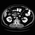
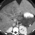
RADIOLOGY: ABDOMEN: Case# 81: NEUROENDOCRINE TUMOR. This 21 year old post-partum black female presents with hyperglycemia and abdominal distention. An large heterogeneously enhancing anterior segment right hepatic lobe mass is identified with central areas of necrosis. A second smaller 5 cm mass is identified within the posterior segment of the right hepatic lobe. Additionally, a large homogeneous pancreatic tail mass is identified. A heterogeneous low attenuation mass with swirled areas of higher attenuation was identified in relation to the inferolateral edge of the pancreatic mass, within the retroperitoneum. Similar abnormal attenuation is identified in the right subhepatic space. The appearance is consistent with new hemorrhage. Caval thrombosis is identified just below the renal veins and extends into the right ovarian vein. There is suggestion of caval thrombosis extension to both iliac veins as well. There is a large amount of ascites. The uterus has typical post-partum appearance. The neuroendocrine tumors of the pancreas are derived from the islet cells. The functioning islet cell tumors include: insulinoma, gastrinoma, glucagonoma, VIPoma, and somatostatinoma. Some islet cell tumors are non-functioning.
- Author
- Peter Anderson
- Posted on
- Thursday 1 August 2013
- Albums
- Visits
- 1588
0 comments