)
)

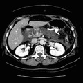
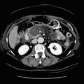
RADIOLOGY: PANCREAS: Case# 77: PANCREATIC NECROSIS. 61-year-old white female with hemorrhagic pancreatitis, status 3 days post exploratory laparoscopic surgery. The pancreatic body and tail are essentially necrosed and replaced by fluid density. Only a minimal amount of enhancing pancreatic tissue remains in the posterior pancreatic neck and head. A percutaneous drain is seen anterior to the pancreatic body and tail fluid density. Inflammatory changes are seen in the pararenal spaces bilaterally with extension into the root of the mesentery. There is mild bowel wall thickening of the colon in the region of the pancreatic bed. Arterial phase scanning through the pancreas can demonstrate those areas within the pancreas that enhance indicating preserved pancreatic parenchyma. Areas which do not enhance and which display liquefaction are likely to be necrotic.
- Author
- Peter Anderson
- Posted on
- Thursday 1 August 2013
- Albums
- Visits
- 1269
0 comments