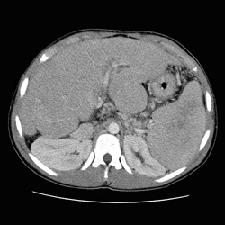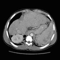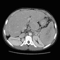
RADIOLOGY: HEPATOBILIARY: Case# 75: PRIMARY BILIARY CIRHOSIS, BILIARY ATRESIA, LIVER MASS. The patient is a 15-year-old male being evaluated for liver transplant for cirrhosis secondary to biliary atresia. Ultrasound performed the previous day demonstrated a mass posterior to the left hepatic lobe. CT is requested for follow-up. The hepatic contour is irregular with enlargement of the left hepatic lobe consistent with a cirrhotic morphology. A lobulated projection off the left lobe posteriorly measuring 4 x 4.5 cm is similar in appearance to the mass described on ultrasound. This lobulation demonstrates homogeneous density similar to the liver. An apparent tissue plane between this lobulated projection and the posterior left hepatic lobe is noted on the inferior images. The liver itself demonstrates moderate heterogeneity with mild dilatation of the intrahepatic ducts confined predominantly to the left hepatic lobe. Pneumobilia is noted in the porta and the right hepatic lobe. Multiple surgical clips are noted just superior to the pancreatic head. The spleen is enlarged demonstrating patchy enhancement on the initial CT images with homogeneous enhancement on delayed scan. Definitive focal splenic lesion is not identified. Multiple perisplenic, gastrohepatic, and mild para-esophageal varices are noted. Mild amount of mesenteric congestion is also seen. There is no evidence of ascites. There is moderate compression of the left kidney by the enlarged spleen, otherwise, the kidneys appear normal. Gallbladder is not seen consistent with patients history of biliary atresia versus prior surgical removal.
- Author
- Peter Anderson
- Posted on
- Thursday 1 August 2013
- Tags
- hepatobiliary, radiology
- Albums
- Visits
- 1184


0 comments