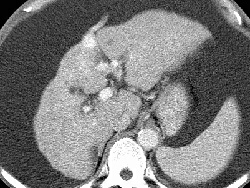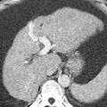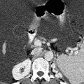
RADIOLOGY: HEPATOBILIARY: Case# 70: ALCOHOLIC CIRRHOSIS W/COLLATERALS. A 65 year old male follow-up case of cirrhosis of the liver. A CT is requested to rule out hepatocellular carcinoma. The liver is small in size with nodular outline. There is relative hypertrophy of the lateral segment of the left lobe as well as caudate lobe. Multiple scattered well-defined round hypodense lesions of varying sizes are noted involving both lobes representing simple hepatic cysts. There is no intrahepatic biliary ductal dilatation. Multiple gallstones are noted incidentally. Massive ascites is seen with multiple collaterals at various locations including paraesophageal, esophageal, paraumbilical, splenic, hilar, gastrosplenic, and gastrohepatic ligament as well as the splenorenal region. There is moderate splenic enlargement without any focal lesions evident. The visualized small as well as large bowel loops are seen floating in the center of mid abdomen. Cirrhosis due to alcoholism usually produces a micronodular pattern. Atrophy of the right lobe with hypertrophy of the left and caudate lobes are also common in alcoholic cirrhosis. Other typical CT findings include fatty infiltration and hepatomegaly, non-homogeneous attenuation, an irregular, nodular contour, decreased liver volume, increased size and prominence of the intrahepatic fissure, ascites, and signs of portal hypertension such as formation of collaterals and splenomegaly.
- Author
- Peter Anderson
- Posted on
- Thursday 1 August 2013
- Tags
- hepatobiliary, radiology
- Albums
- Visits
- 1247


0 comments