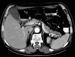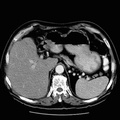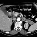
RADIOLOGY: GASTROINTESTINAL: GI: Case# 63: EXTRA-PANCREATIC GASTRINOMA. A 64 year old male with a six month history of abdominal pain and forty pound weight loss. Zollinger-Ellison syndrome is a diagnostic consideration. There is marked soft tissue density thickening along the fundus of the stomach, which extends up into the gastroesophageal junction. The etiology of this finding is uncertain. No intraparenchymal pancreatic mass is identified. A small mass is seen medial to the pancreatic head adjacent to the duodenum. Another tiny mucosal nodule was identified retrospectively in the lateral duodenal wall. A nuclear medicine octreotide scan performed soon after the CT scan was positive in the region of the duodenum adjacent to the pancreatic head. Non-functioning islet-cell tumors can grow to be quite large before clinical detection. Functioning islet-cell tumors, however, are usually small when clinical findings become evident. In the case of gastrinoma, the findings of Zollinger-Ellison syndrome may be apparent: excess acid production with gastric/duodenal inflammation and/or multiple peptic ulcers. Most islet-cell tumors are found within the parenchyma of the pancreas. Occasionally, however, they may be discovered in an ectopic location: stomach, duodenum, jejunum, or splenic hilum. CT usually cannot detect islet-cell tumors in these ectopic locations.
- Author
- Peter Anderson
- Posted on
- Thursday 1 August 2013
- Tags
- gastrointestinal, radiology
- Albums
- Visits
- 1538


0 comments