)
)

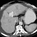
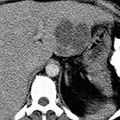
RADIOLOGY: HEPATOBILIARY: Case# 56: CIRRHOSIS, HCC. This is a 68 year old male with cirrhosis who now presents with elevated amylase, lipase as well as white blood cell count. CT scan of the abdomen is requested to rule out abscess and/or hepatic cell carcinoma. The liver is nodular and has a slightly lower attenuation than that of the spleen. A moderate amount of ascites is seen to surround the liver and spleen extending to the pelvis. A recannalized umbilical vein is noted to drain into the left portal vein. All four segments of the liver contain low attenuation lesions. The largest of these is in the lateral segment of the left lobe of the liver and measures 3.2 x 1.8 cm. A heterogeneous 5 x 6 cm ill-defined region is noted in the medial segment of the left lobe of the liver adjacent to the falciform ligament; this lesion extends into the anterior segment of the right lobe. Additional low attenuation regions are also noted in the posterior segment of the right lobe of the liver as well as a region at the dome of the lateral segment of the left lobe of the liver. There is no intrahepatic biliary ductal dilatation. The right portal vein does not enhance, consistent with occlusion. The gallbladder wall is mildly thickened. The spleen appears unremarkable. Although uncommon in the United States, hepatocellular carcinoma is an important cause of death in parts of Africa and Asia because of the hepatotrophic viruses. In the United States, eighty percent of hepatocellular carcinomas arise in cirrhotic livers. Three patterns of tumor growth are seen: 50% present as a solitary tumor, 30% as a diffuse infiltrative tumor, and 20% as a multinodular tumor. The tumor usually appears as a hypodense or isodense lesion on nonenhanced images and enhances prominently during the arterial phase on dynamic contrast injection. Areas of tumor necrosis and calcification are common. Tumor invasion of hepatic and portal veins occurs frequently. Metastases usally involve lung, regional nodes or occasionally bone.
- Author
- Peter Anderson
- Posted on
- Thursday 1 August 2013
- Tags
- hepatobiliary, radiology
- Albums
- Visits
- 1442
0 comments