)
)

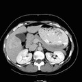
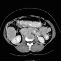
RADIOLOGY: HEPATOBILIARY: Case# 52: HEPATIC MASSES. Evaluate hepatic hemangiomas. Two focal abnormalities are seen within the liver. The first, in the anterior segment of the right lobe, measures 5 x 5 cm. On the noncontrast studies, this is moderately well-circumscribed and is hypodense. Following contrast, there is peripheral enhancement of this lesion and on delayed images the lesion is isodense with hepatic parenchyma. The second lesion measures 7 x 4.7 cm and arises from the tip of the right lobe. This lesion shows less convincing peripheral enhancement and is also isodense on delayed on images. No other hepatic lesion is seen. The pancreas, gallbladder, spleen, adrenals, and kidneys are normal. There is no lymphadenopathy or free intraperitoneal fluid. The uterus and ovaries are normal. Lung bases and bones are clear.
- Author
- Peter Anderson
- Posted on
- Thursday 1 August 2013
- Tags
- hepatobiliary, radiology
- Albums
- Visits
- 1339
0 comments