)
)

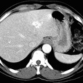
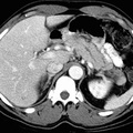
RADIOLOGY: HEPATOBILIARY: Case# 51: HEMANGIOMAS - 3 PHASE. The patient is a 53 year old male to rule out hepatocellular carcinoma. Three hemangiomas are identified: A 2 cm nodular lesion in the lateral segment of the left hepatic lobe. Approximately 1 cm lesion in the anterior segment of the right hepatic lobe near the dome of the liver . Approximately 1 cm lesion at the anterior aspect of the right hepatic lobe near the periphery of the liver . These show early nodular, peripheral enhancement and progressive filling in on venous phase images. The pancreas is mildly enlarged and heterogeneous in appearance with a dilated pancreatic duct. Hemangiomas are a common incidental finding on routine imaging surveys of the liver and may pose an important diagnostic problem in patients with a known primary malignancy. Hemangiomas are the most common benign tumors of the liver. They are usually found in the right hepatic lobe and are subcapsular in location. The CT appearance of hemangiomas after a bolus injection of iodinated contrast material with dynamic scanning demonstrates a dense peripheral enhancement, which eccentrically fills in toward the center of the lesion on delayed scan. This is the most common finding and is seen in approximately 85% of lesions greater than 3cm in size. The angiographic appearance of a hemangioma is classic, with puddling of contrast material within the tumor, which remains visible on delayed, prolonged filming sequences. Some hemangiomas exhibit atypical features such as cystic areas probably caused by hemorrhage or thrombosis. Other lesions simply fail to show the typical pattern well enough to confirm the diagnosis.
- Author
- Peter Anderson
- Posted on
- Thursday 1 August 2013
- Tags
- hepatobiliary, radiology
- Albums
- Visits
- 1272
0 comments