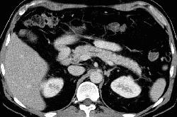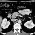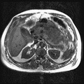
RADIOLOGY: PANCREAS: Case# 48: SMALL PANCREATIC HEAD MASS-ACINAR ATROPHY, STROMAL FIB., CHRONIC INFLAM. (NEGATIVE FOR MALIGNANCY). 70 year old male with possible pancreatic head mass on previous CT. He reports weight loss and anorexia. A recent abdominal CT performed at an outside institution is reviewed. A 24 x 15mm mass with subtle central low attenuation is present in the uncinate process of the pancreas. It is separate from the mesenteric vessels and the inferior vena cava, however, it is not separable from the medial wall of the second portion of the duodenum. There is minimal dilatation of the common bile duct and intrahepatic biliary system proximal to this mass. The pancreatic duct is slightly prominent but not actually dilated.
- Author
- Peter Anderson
- Posted on
- Thursday 1 August 2013
- Albums
- Visits
- 1785


0 comments