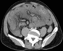
RADIOLOGY: VASCULAR: Case# 43: H/O HOMOCYSTINURIA, PV & SMV THROMBOSIS. 27-year-old gentleman with upper and lower GI tract bleeding and ahistory of homocystinuria. The portal vein is enlarged with lack of enhancement in the main, right and left branches. The superior mesenteric vein and splenic vein are also enlarged markedly with diminished enhancement. A moderate amount of free intraperitoneal fluid is identified. There is patchy enhancement diffusely throughout the liver and spleen, probably on the basis of poor perfusion. Throughout the small bowel, the folds are markedly thickened, though the loops themselves are not dilated. Thrombosis of the portal vein may be caused by neoplasm, infection, trauma, hypercoaguable states, or hepatic venous obstruction. In the acute phase, the portal vein is enlarged and has a CT attenuation value equal to that of unopacified blood on noncontrast images; it does not demonstrate central enhancement with contrast. Rarely, the thrombus may be of higher attenuation which is typical of freshly clotted blood. There may be segmental or focal decrease in hepatic enhancement to the segment(s) supplied by the occluded portal vein branches. The course of the thrombus may be seen extending into the splenic or superior mesenteric veins.