)
)

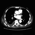
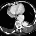
RADIOLOGY: AORTA: Case# 27: LEAKING ABDOMINAL AORTIC ANEURYSM. 84-year-old male with a known abdominal aortic aneurysm who presents with severe abdominal pain. There is a very large abdominal aortic aneurysm which begins between the origin of the superior mesenteric artery and the renal artery and extends into the aortic bifurcation to involve the right common iliac artery. At its greatest dimensions, it measures 15.0 cm x 10.0 cm and contains a large amount of intramural thrombus. The aortic aneurysm is eccentric in location, residing more in the right side of the retroperitoneum. There is a moderate amount of hemorrhage within the right side of the retroperitoneum surrounding the aneurysm consistent with subacute hemorrhage. No active arterial extravasation is identified at this time. Abdominal aortic aneurysms usually occur in the setting of atherosclerotic disease but may be caused by syphilis, by extension of a dissection from above, or by connective tissue disorders such as Takayasus arteritis. Complications of abdominal aortic aneurysm include rupture, peripheral embolization, thrombosis, and infection. The incidence of rupture increases with increasing aneurysm size above 4cm. Rupture usually occurs into the left retroperitoneal space and rarely into the gastrointestinal tract and inferior vena cava. If a ruptured or leaking aneurysm is suspected, CT should be done with contrast enhancement. Typical findings include obscuration or anterior displacement of the aneurysm by an irregular high density mass or collection that extends into one or both perirenal spaces. The wall of the aneurysm may be identified by calcifications while the lumen enhances. Other findings include anterior displacement of the kidney by hematoma, enlargement or obscuration of the psoas muscle, and a focally indistinct aortic margin that corresponds to the site of rupture. In contrast, a chronic pseudoaneurysm appears as a well-defined, ususally round mass with attenuation similar or lower than that of the native aorta on noncontrasted images.
- Author
- Peter Anderson
- Posted on
- Thursday 1 August 2013
- Albums
- Visits
- 2028
0 comments