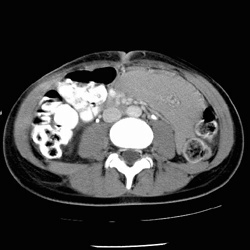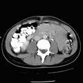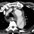
RADIOLOGY: VASCULAR: Case# 25: MESSENTERIC HEMATOMA. Fourteen year old white female 2 days status post MVA as a restrained passenger. A complex intrahepatic laceration is seen to predominantly involve the anterior segment of the right lobe with extension into the porta. The laceration does not appear to extend to the liver surface. Only a small amount of fluid is seen around the liver. In the left mid abdomen, there is a high attenuation interlooped collection which most likely represents a hematoma. Well-opacified bowel loops are seen coursing through this collection. Mesenteric hematomas may be caused by blunt trauma to the abdomen, excessive anticoagulation or postoperative bleeding. On CT scan, a mesenteric hematoma appears as a fluid collection within the mesentery of the bowel. A relatively high attenuation value indicates fresh blood within the hematoma.
- Author
- Peter Anderson
- Posted on
- Thursday 1 August 2013
- Albums
- Visits
- 2381


0 comments