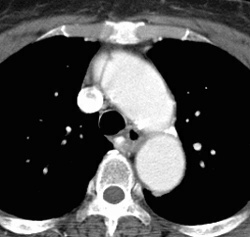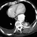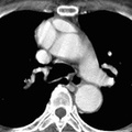
RADIOLOGY: AORTA: Case# 22: TYPE A AORTIC DISSECTION. 70 year old female with chest pain. A linear filling defect is present in the proximal ascending aorta, representing an intimal flap dividing a true and false lumen. More superiorly, a second false lumen is seen. The intimal flaps remain proximal to the take off of the left subclavian artery. Aortic dissection results from a tear in the vessel wall with leakage of blood into the media creating a false channel or lumen. This false channel is typically between the inner one third and outer two thirds of the media. Most cases of dissection are due to intrinsic weakness of the media secondary to a connective tissue disorder or arteritis. Less commonly, arteriosclerosis or syphilis may predispose to dissection.
- Author
- Peter Anderson
- Posted on
- Thursday 1 August 2013
- Albums
- Visits
- 1846


0 comments