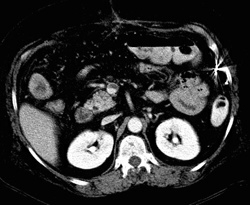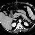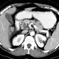
RADIOLOGY: VASCULAR: Case# 20: SMV & PV THROMBOSIS, BOWEL EDEMA, ITP. 54 yo female with ITP and hepatitis who presents with severe abdominal pain, nausea and vomiting. There is thrombosis of the right and left portal veins, main portal vein, splenic vein and superior mesenteric vein. Thrombus extends into a segmental branch of the SMV (seen on the last image). In the portion of bowel drained by the thrombosed SMV, the bowel wall is thickened. Diffuse strandy inflammatory changes are also apparent in the region of ileum and ascending colon indicating mesenteric congestion. SMV thrombosis can be an acute or chronic process. In acute SMV thrombosis, the SMV may become enlarged and have a high attenuation value (equal to or higher than soft tissue). Chronic SMV thrombosis is characterized by mild enlargement of the vein with central low denstiy surrounded by higher density wall. Associated findings may include increased attenuatiuon of the mesenteric fat due to mesenteric edema and bowel wall thickening due to stasis and mesenteric ischemia.
- Author
- Peter Anderson
- Posted on
- Thursday 1 August 2013
- Albums
- Visits
- 4009


0 comments