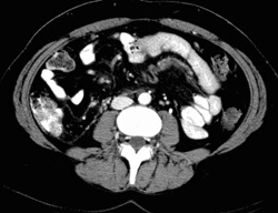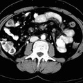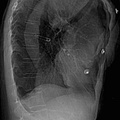
RADIOLOGY: VASCULAR: Case# 18: SMV THROMBOSIS. This is a 43 year old female who has fever, abdominal pain, nausea and vomiting as well as elevated LFTs. Ultrasonography revealed thrombus in the porto-splenic confluence extending into the SMV and main portal vein. The liver parenchyma appears unremarkable. The main portal vein near the pancreatic head has a low attenuation region centrally, consistent with non-occlusive thrombus. This thrombus becomes occlusive at the portal confluence and completely occlusive thrombus extends into the superior mesenteric vein to the level of the umbilicus. Additionally, the mesenteric root surrounding the superior mesenteric vein displays strandy changes consistent with inflammation. Etiologies of thrombosis of the SMV are similar to those in portal vein thrombosis. Cirrhosis with portal hypertension impairs mesenteric venous flow leading to stasis. Extrinsic compression by neoplasm, especially pancreatic, may produce thrombosis. Other processes which may induce thrombosis are blunt trauma, inflammatory processes such as pancreatitis or peritonitis, and hypercoaguable states. Approximately one-half of cases of SMV thrombosis are idiopathic.
- Author
- Peter Anderson
- Posted on
- Thursday 1 August 2013
- Albums
- Visits
- 2433


0 comments