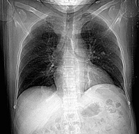
RADIOLOGY: CHEST: Case# 13: LIPOSARCOMA R SUBCLAVIAN AND SVC. 45 year old male with a history of atrial fibrillation and a one year history of right anterior chest wall pain. Patient recently had a cardiac catheterization which did not show any significant coronary artery disease. There is an abnormal fat collection at the right lung apex that seems to engulf the right subclavian vein and either expands the right brachiocephalic vein or compresses and flattens it. Extensive collaterals are noted around the right shoulder and posterior chest wall with dense opacification of the azygous vein. However, the superior vena cava is patent. Liposarcoma is the second most common soft tissue sarcoma (after malignant fibrous histiocytoma) with the bulk of the tumor differentiating into adipose tissue. This tumor is common in 5-6th decade with 40-50% being myxoid type. These may be painful in 10-15% of cases and more common in the lower extremity (41%) and trunk (42%). The tumor may not be well seen on routine radiographs due to the fat content but CT shows inhomogenous mass with fat content. Charecteristic enhancement after contrast administration distinguishes this tumor from a lipoma. Concomitant lesions may be seen in other areas in 10%.