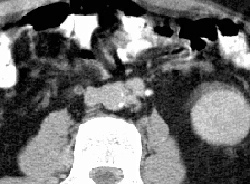
RADIOLOGY: VASCULAR: Case# 12: CHRONIC PTE AND RT DVT. 49-year old man with h/o sudden onset SOB. Doppler ultrasound examination of the lower extremities revealed femoral vein thrombus. VQ scan showed complete lack of perfusion of the right lung. A markedly dilated azygous vein is identified. Also, dilated azygous and hemiazygous veins are identified in the retrocrural space. There is acute thrombus within the common iliacs, external and internal iliac veins, and common femoral veins bilaterally. Above the confluence of the common iliac veins, the inferior vena cava is inapparent. Multiple collateral vessels are present around the aorta and the azygous and hemiazygous veins are prominent. The portal venous axis is patent. Over 600,000 cases of pulmonary embolism occur each year in the United States, with one-third of episodes leading to death. Findings are nonspecific and often lead to a failure to diagnose. The classic triad of dyspnea, pleuritic chest pain, and hemoptysis is seen in only 20% of patients with PE. The mortality rate for untreated PE is 30% versus 8% for treated PE. The most common source for pulmonary emboli is deep venous thrombosis of the lower extremities. Other sites include the internal iliac veins, the inferior vena cava, upper extremities, mural thrombus within the right heart, renal veins and tumor emboli. Ventilation and perfusion scintography and pulmonary arteriography are used to diagnose PE. CT may be used in patients who are at high risk for complications of arteriography. On CT, emboli appear as intraluminal filling defects. There may be evidence of infarction, such as wedge-shaped areas of parenchymal consolidation adjacent to a pleural surface. Linear strands may extend from the lesion toward the hilum, and the periphery of the lesion may enhance after intravenous administration of contrast, probably due to collateral flow. In cases of massive PE without infarction, the oligemic lobe may appear normal while the normal areas may look consolidated. Atelectasis and pleural effusion are often present.