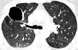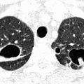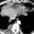
RADIOLOGY: LUNG: Case# 10: PULMONARY ASPERGILLOMA, S/P BMT. 47 year old patient status post bone marrow transplantation for leukemia. A large cystic lesion containing soft tissue nodular opacity is identified in the posterior segment of the right upper lobe. A small cavitary lesion with a fungus ball is also identified in the posterior segment of the left upper lobe. Cavitary lesions are seen in the superior segment of the left lower lobe and in the anterior segment of the right upper lobe. No pleural effusion is identified, and the mediastinal soft tissue windows do not show any enlarged lymph nodes. Aspergillus produces several different clinical illnesses in humans: acute bronchopulmonary aspergillosis which is an IgE hypersensitivity reaction presenting with symptoms similar to asthma, and aspergilloma or a fungus ball, which may present as hemoptysis. Aspergillus is important in immunosuppressed individuals due to the possibility of invasive pulmonary aspergillosis. Fungus balls usually form within pre-existing pulmonary cavities from TB, old infarcts or abscesses, and bronchiectasis. Early in the formation of an aspergilloma, an irregular spongelike network filling the cavity may be evident on CT. Other findings include a pulmonary mass surrounded by a zone of lower attenuation due to edema (the halo sign) and associated pleural thickening.
- Author
- Peter Anderson
- Posted on
- Thursday 1 August 2013
- Albums
- Visits
- 2130


0 comments