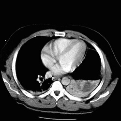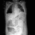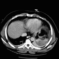
RADIOLOGY: CHEST: Case# 4: L. DIAPHRAGMATIC RUPTURE. The patient was a restrained driver of a vehicle struck on the passenger side in an MVA. The stomach is seen in an intrathoracic position. Complete atelectasis of the basilar segments of the left lower lobe are noted, presumably due to the hernia. No pneumothorax is seen. Dependent atelectasis is seen in the right lung base. Two rib fractures are noted in the left posterior chest at the level of the superior aspect of the spleen. Chest radiograph is the principal method of screening for intrathoracic injury after blunt abdominal trauma. Chest radiographs have a sensitivity of 45% on left and 17% on right and 27% both sides combined, in diagnosing diaphragmatic rupture (DR). If chest radiograph suggests the diagnosis a CT scan is generally not needed for the purpose of diagnosing DR. However if Chest X-ray is negative CT scan has a sensitivity of 60% and specificity of 87%, the most useful and commonly seen sign being loss of continuity of the diaphragm. Other signs are visceral herniation, "collar sign", and presence of both hemoperitoneum and hemothorax.
- Author
- Peter Anderson
- Posted on
- Thursday 1 August 2013
- Albums
- Visits
- 2469


0 comments