File list
This special page shows all uploaded files.
| Date | Name | Thumbnail | Size | Description | Versions |
|---|---|---|---|---|---|
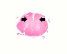
| 02:43, 21 August 2013 | IPLab8Polio1.jpg (file) |  |
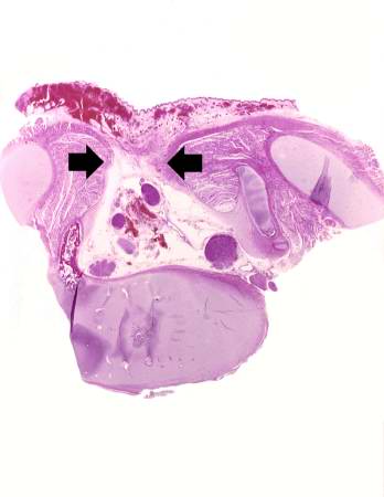
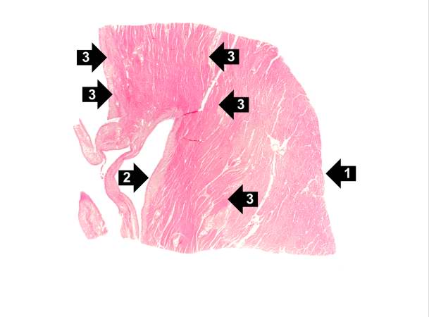
3 KB | This is a low-power photomicrograph of a section of spinal cord from this case. Note that the anterior horns (arrows) are almost completely devoid of neurons. | 1 |
| 02:13, 21 August 2013 | IPLab7Osteosarcoma3.jpg (file) |  |
10 KB | This is another view of the tumor in the distal femur. | 1 |
| 15:12, 15 August 2013 | IPLab1KidneyInfarction3.jpg (file) |  |
11 KB | 1 | |
| 18:07, 19 August 2013 | IPLab6RA5.jpg (file) |  |
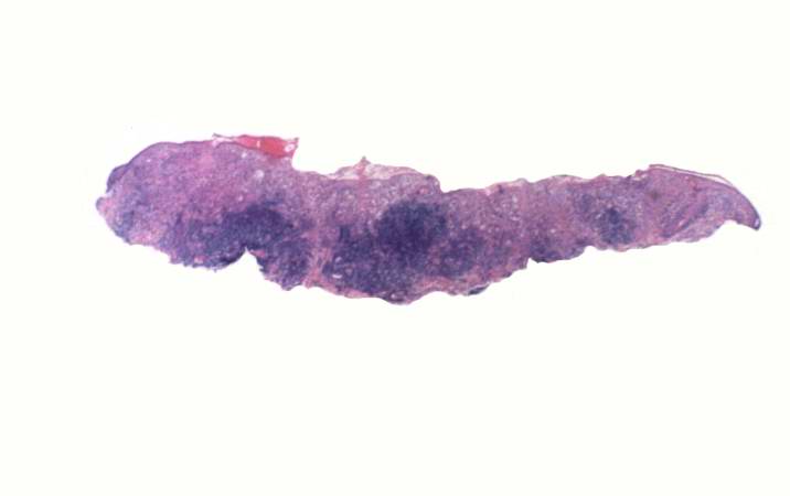
11 KB | This is a low-power photomicrograph of the subcutaneous nodule from this patient. | 1 |
| 01:50, 21 August 2013 | IPLab7IDC2.jpg (file) |  |
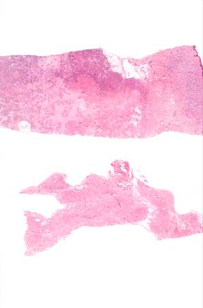
12 KB | These are sections of normal breast (lower) and breast tissue with infiltrating duct carcinoma (upper). Note the increased cellularity (increased blue staining due to the increased number of nuclei) in the tumor tissue. | 1 |
| 02:12, 19 August 2013 | IPLab3AcuteAppendicitis7.jpg (file) |  |
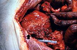
13 KB | This is a gross photograph of the open abdominal cavity of a patient with acute appendicitis. In this patient, there had been rupture of the appendix with spillage of intestinal contents into the abdominal cavity. This spillage resulted in an acute abd... | 1 |
| 02:10, 19 August 2013 | IPLab3AcuteAppendicitis2.jpg (file) |  |
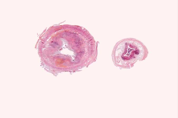
13 KB | This is a low-power photomicrograph of a normal appendix on the right and an appendix with acute inflammatory response on the left. Note the abundant blue-stained lymphoid tissue beneath the mucosal layer and the absence of blue-staining cells in the s... | 1 |
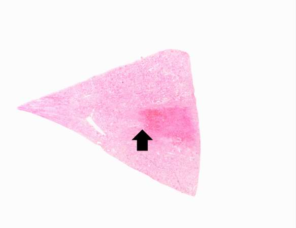
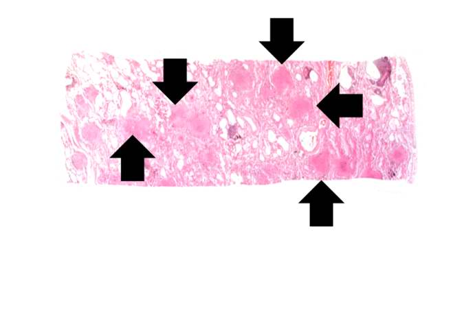
| 04:06, 21 August 2013 | IPLab10Histo1.jpg (file) |  |
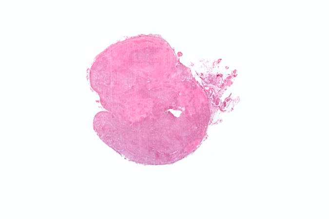
13 KB | This low-power photomicrograph shows a section of adrenal gland with several irregularly-outlined areas of necrosis. | 1 |
| 05:04, 21 August 2013 | IPLab11Cysticercosis4.jpg (file) |  |
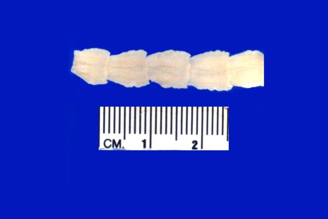
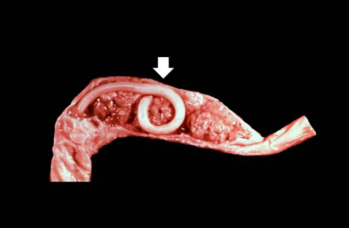
13 KB | This is a photograph of the body of an adult tapeworm. Note the body segments. | 1 |
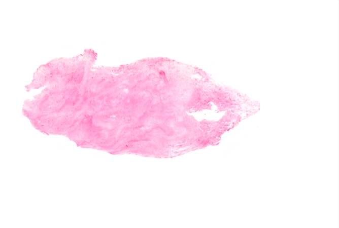
| 04:58, 21 August 2013 | IPLab11Leishmaniasis2.jpg (file) |  |
13 KB | This is a low-power photomicrograph of the biopsy taken from this skin lesion. The ulcerated surface is at the top. Note that the specimen is heavily infiltrated with inflammatory cells. | 1 |
| 04:10, 19 August 2013 | IPLab3FibrinousPericarditis3.jpg (file) |  |
13 KB | This low-power photomicrograph illustrates the dark-red-staining fibrin deposits on the inner surface (arrows). This pericardium is much thicker than normal and there are numerous inflammatory cells within the pericardial tissue. | 1 |
| 05:44, 21 August 2013 | IPLab13Myelomeningocele2.jpg (file) |  |
15 KB | This gross photograph shows consecutive lumbar vertebra from this case. Note the defect (arrows) in the two vertebral bodies on the right. This defect was caused by failure of the vertebral column to properly close. | 1 |
| 05:45, 21 August 2013 | IPLab13Myelomeningocele4.jpg (file) |  |
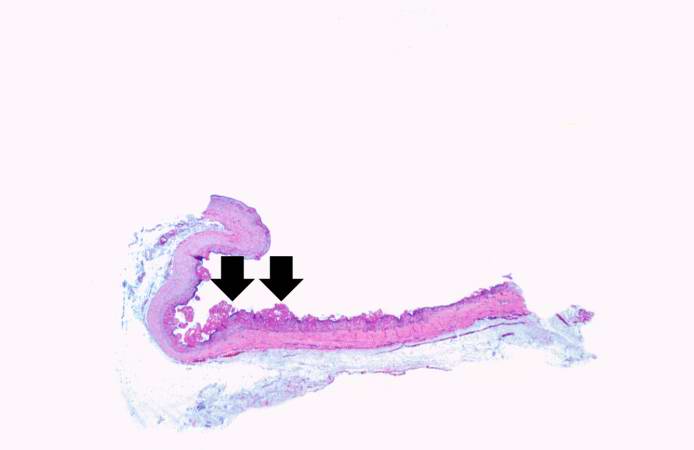
16 KB | This is a low-power photomicrograph of one of the vertebral bodies from this case. In this section there are defects (arrows) in the vertebral body but the skin can be seen over the open vertebral canal. | 1 |
| 05:01, 21 August 2013 | IPLab11Ascariasis2.jpg (file) |  |
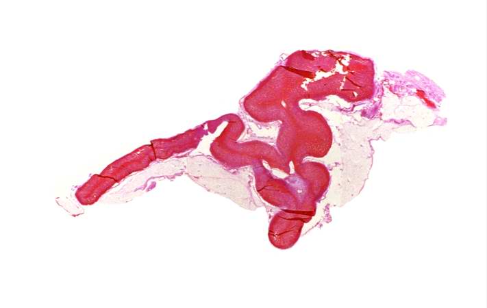
16 KB | This is a photograph of an autopsy specimen from another case of ascariasis. The adult ascarid (arrow) can be seen in the section of small bowel from this patient. | 1 |
| 05:45, 21 August 2013 | IPLab13Myelomeningocele3.jpg (file) |  |
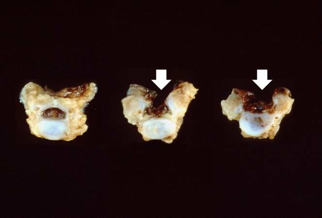
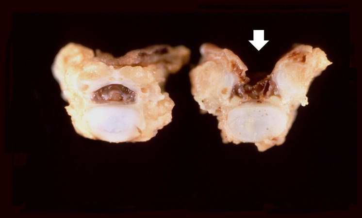
17 KB | This is a closer view of the previous gross photograph showing a normal lumbar vertebra from this case on the left. Once again, note the defect (arrow) in the vertebral body on the right due to failure of the vertebral column to close properly. | 1 |
| 18:27, 19 August 2013 | IPLab5Downs2.jpg (file) |  |
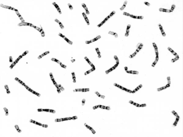
17 KB | These cells, obtained by amniocentesis, were cultured and then arrested in metaphase. Nuclei from these cells were isolated and stained to demonstrate the banding pattern of each chromosome. This photograph shows a "chromosome spread." Each chromosome ... | 1 |
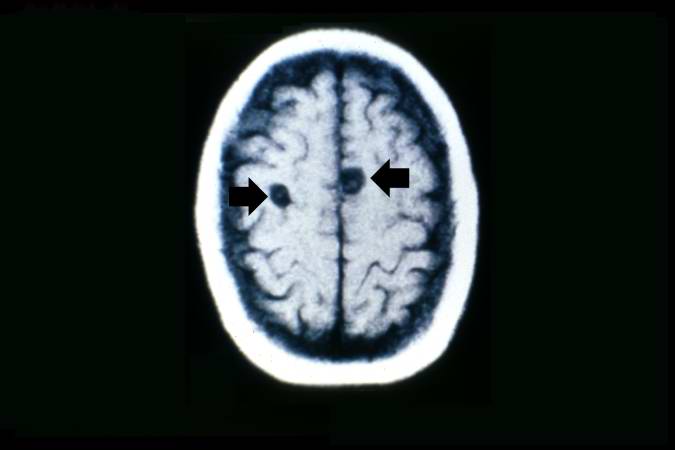
| 05:03, 21 August 2013 | IPLab11Cysticercosis1.jpg (file) |  |
17 KB | This is a head CT showing the two cysts (arrows). | 1 |
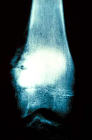
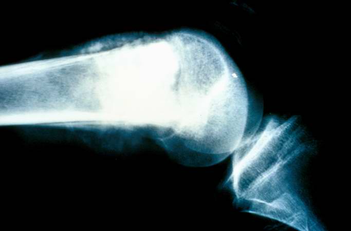
| 02:13, 21 August 2013 | IPLab7Osteosarcoma2.jpg (file) |  |
17 KB | This is a radiograph showing the tumor in the distal femur. | 1 |
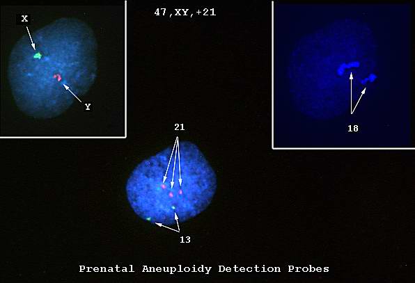
| 18:27, 19 August 2013 | IPLab5Downs1.jpg (file) |  |
18 KB | This is a photomicrograph of cells obtained by amniocentesis that were stained using FISH. The cell in panel 1 was stained with markers specific for the X and Y-chromosomes. The cell in panel 2 was stained with a marker specific for chromosome 18. The ... | 1 |
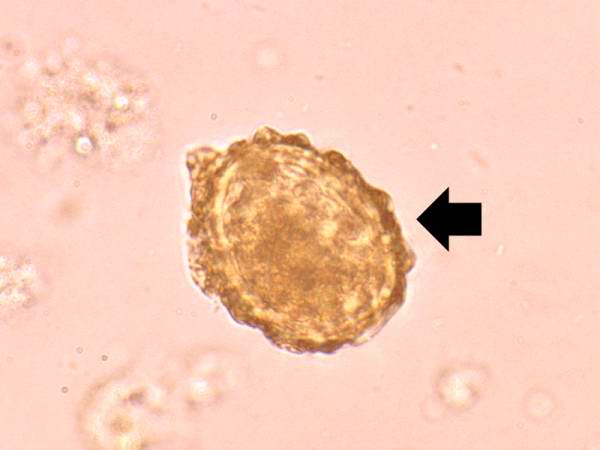
| 05:01, 21 August 2013 | IPLab11Ascariasis3.jpg (file) |  |
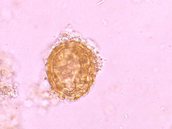
19 KB | This is a high-power photomicrograph of a fecal specimen from this patient showing an ascarid egg (arrow). | 1 |
| 04:58, 21 August 2013 | IPLab11Leishmaniasis6.jpg (file) |  |
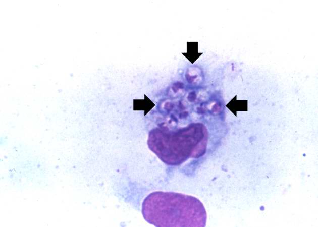
19 KB | This is a high-power photomicrograph of a touch prep made from the skin lesion at the time of biopsy. A single macrophage can be seen with intracytoplasmic leishmania organisms (arrows). | 1 |
| 02:13, 21 August 2013 | IPLab7Osteosarcoma1.jpg (file) |  |
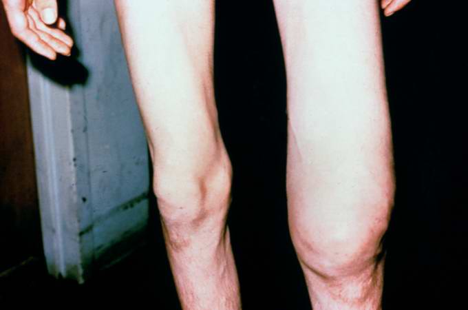
19 KB | This is a photograph of the patient prior to surgery. Note the marked swelling of the knee. | 1 |
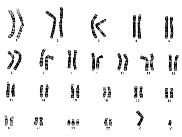
| 18:28, 19 August 2013 | IPLab5Downs5.jpg (file) |  |
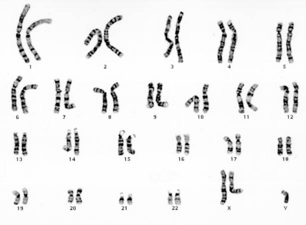
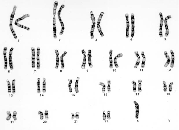
19 KB | This is a karyotype of a patient with Klinefelter syndrome (47, XXY). | 1 |
| 06:05, 21 August 2013 | IPLab13Meningococcemia6.jpg (file) |  |
20 KB | This is a low-power photomicrograph of the adrenal gland from this case. Note that the entire gland is hemorrhagic. | 2 |
| 05:03, 21 August 2013 | IPLab11Cysticercosis2.jpg (file) |  |
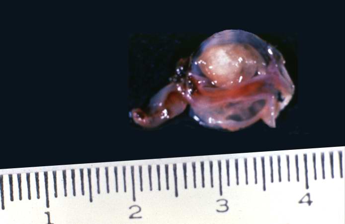
20 KB | This is a photograph of the cyst that was surgically removed. The cyst is filled with a clear fluid and contains a scolex. | 1 |
| 18:29, 19 August 2013 | IPLab5Downs6.jpg (file) |  |
20 KB | This is a karyotype of a patient with Turner syndrome (45, X). | 1 |
| 16:22, 19 August 2013 | IPLab4ChronicPassiveCongestion8.jpg (file) |  |
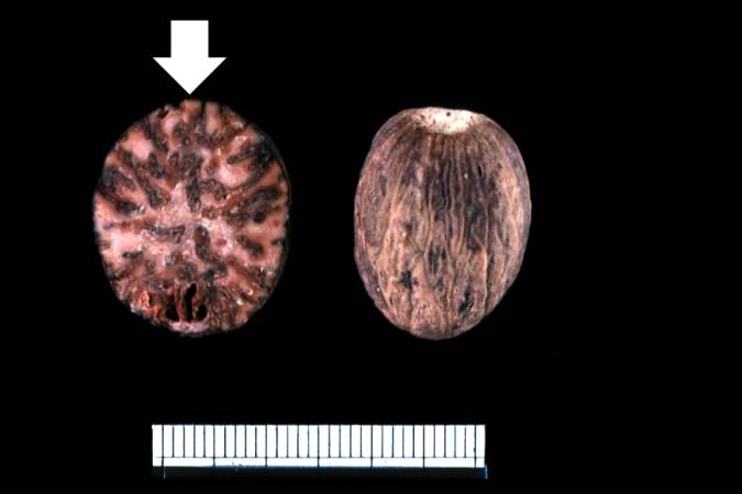
21 KB | This is a gross photograph of a nutmeg. You can see from the appearance of the cut surface of the nutmeg (arrow) why chronic passive congestion of the liver is sometimes referred to as "nutmeg liver." | 1 |
| 01:55, 21 August 2013 | IPLab7Melanoma3.jpg (file) |  |
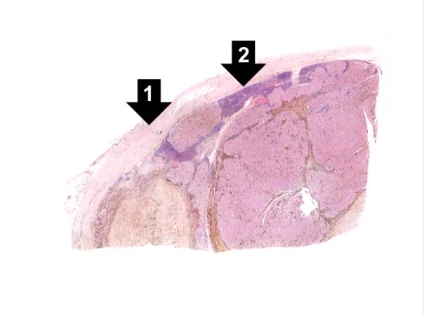
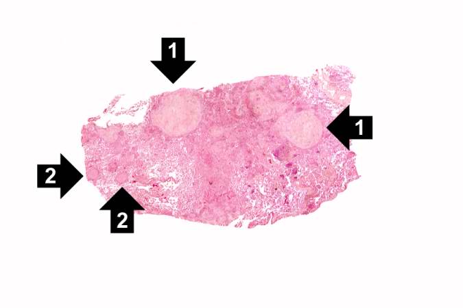
21 KB | This is a low-power photomicrograph of lymph node that is almost completely replaced/filled with tumor. This lymph node has a capsule (1) and some remaining lymphocytes (2) but the remainder of the node is replaced by tumor cells. | 1 |
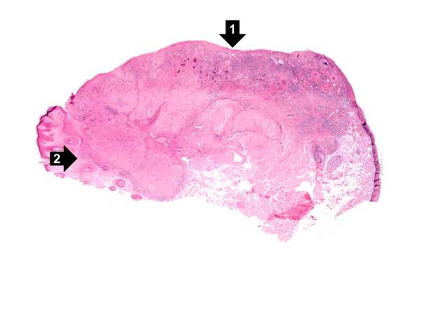
| 01:31, 21 August 2013 | IPLab7LipSCC2.jpg (file) |  |
21 KB | This is a low-power photomicrograph of squamous cell carcinoma of the lip. Note focal ulceration (1) and tumor infiltration at the vermilion border (2). | 1 |
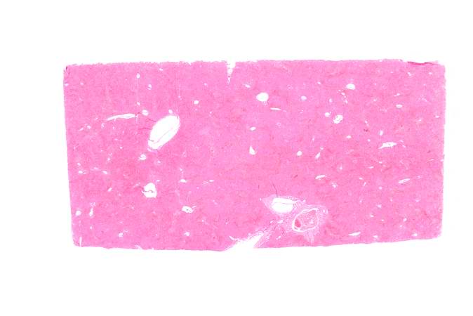
| 16:20, 19 August 2013 | IPLab4ChronicPassiveCongestion3.jpg (file) |  |
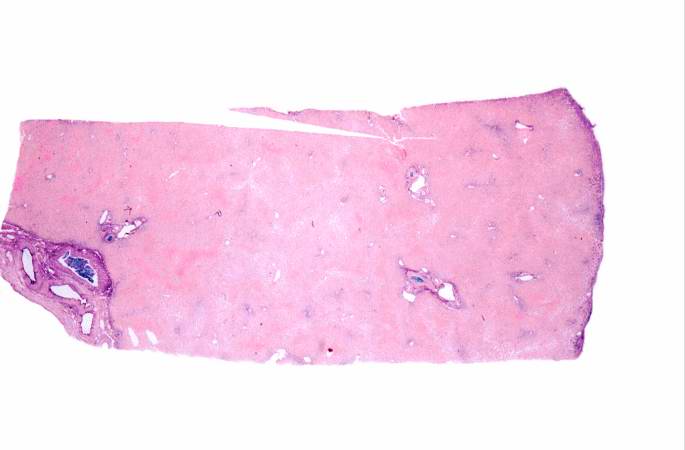
21 KB | This low-power photomicrograph of liver demonstrates a slightly visible pattern of centrilobular congestion at this magnification. | 1 |
| 18:28, 19 August 2013 | IPLab5Downs3.jpg (file) |  |
21 KB | Chromosomes from the chromosome spread are lined up to demonstrate the karyotype. In this case there are three copies of chromosome 21, just as noted in the FISH. | 1 |
| 02:50, 16 August 2013 | IPLab1Tuberculosis4.jpg (file) |  |
21 KB | 1 | |
| 06:06, 21 August 2013 | IPLab13Meningococcemia8.jpg (file) |  |
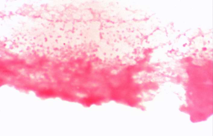
21 KB | This is a higher-power photomicrograph of a smear of cerebrospinal fluid taken at autopsy. Note the Gram-negative cocci in this smear, indicative of N. meningitidis. | 1 |
| 04:55, 21 August 2013 | IPLab11Malaria3.jpg (file) |  |
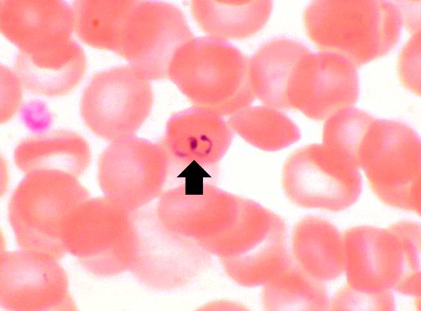
22 KB | This is yet another high power photomicrograph of a thin smear of blood from this patient. There is one RBC that contains two ring stage trophozoites (arrow). This is characteristic of, but not diagnostic for, P. falciparum. | 1 |
| 02:47, 21 August 2013 | IPLab8HBV1.jpg (file) |  |
22 KB | This is a low-power photomicrograph of liver from this case. This section was stained with a modified aldehyde fuchsin and counterstained with hematoxylin and eosin. Modified aldehyde fuchsin colors cystine-rich proteins--such as HBsAg and elastic fibe... | 1 |
| 13:49, 15 August 2013 | IPLab1MyocardialInfarction2.jpg (file) |  |
22 KB | 1 | |
| 03:38, 19 August 2013 | IPLab3Tuberculosis2.jpg (file) |  |
22 KB | This low-power photomicrograph of a section of lung reveals multiple large nodules (1) with pale eosinophilic centers surrounded by a rim of blue-staining nuclei. In addition to the large nodules, there are several smaller nodules throughout the slide ... | 1 |
| 05:01, 21 August 2013 | IPLab11Ascariasis4.jpg (file) |  |
22 KB | This is a higher-power photomicrograph of another ascarid egg. | 1 |
| 18:28, 19 August 2013 | IPLab5Downs4.jpg (file) |  |
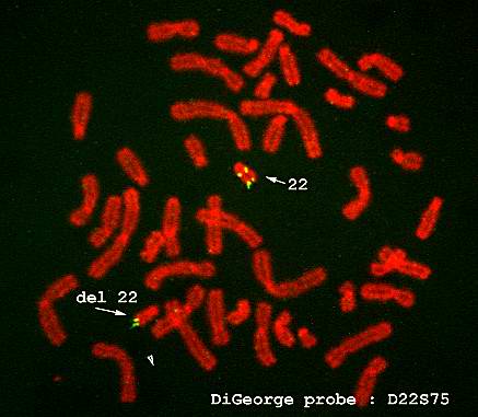
22 KB | FISH is also useful in the diagnosis of other genetic disorders. This is an example of FISH staining on another patient using a probe specific for DiGeorge's disease. The arrow shows that there is a deletion on chromosome 22, which is diagnostic for Di... | 1 |
| 02:49, 21 August 2013 | IPLab8HBV7.jpg (file) |  |
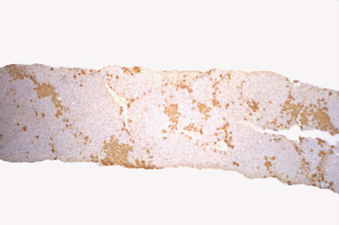
23 KB | This is a low-power photomicrograph of liver from the previous image which has been reacted with antibody specific for HBsAg. The hepatocytes that contain HBsAg stain brown. | 1 |
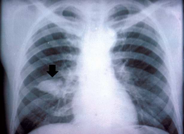
| 04:10, 21 August 2013 | IPLab10Crypto1.jpg (file) |  |
23 KB | This is the chest x-ray showing the mass (arrow) in the right lower lobe. | 1 |
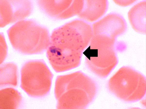
| 04:56, 21 August 2013 | IPLab11Malaria7.jpg (file) |  |
24 KB | In this peripheral smear from a different patient who was infected with P. vivax, the cytoplasm of the infected RBC has a stippled appearance (Schüffner's dots) (arrow). The RBC is also slightly enlarged. | 1 |
| 18:06, 19 August 2013 | IPLab6RA4.jpg (file) |  |
24 KB | This is a gross photograph of the foot from this same patient. Note the subcutaneous nodule on the medial aspect of the foot (arrow). | 1 |
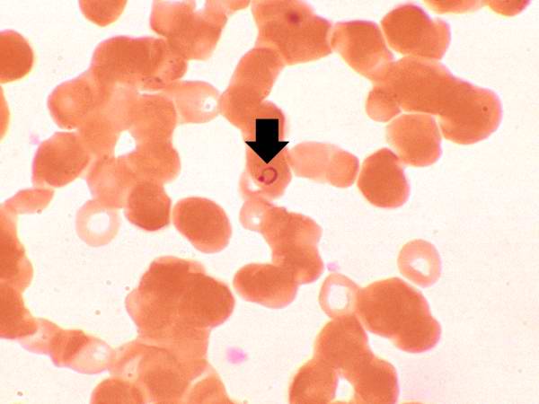
| 04:54, 21 August 2013 | IPLab11Malaria1.jpg (file) |  |
24 KB | This is a high power photomicrograph of a thin smear of blood from this patient. Note that one of the RBCs has a ring stage trophozoite (arrow). | 1 |
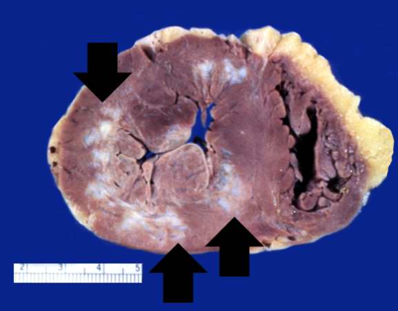
| 04:36, 19 August 2013 | IPLab3HealedMyocardialInfarction1.jpg (file) |  |
25 KB | This is a gross photograph of a heart with areas of old healed myocardial infarction (scars) outlined by arrows. | 1 |
| 02:39, 21 August 2013 | IPLab8Rabies1.jpg (file) |  |
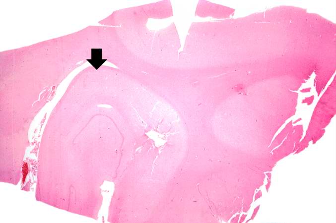
25 KB | This is a low-power photomicrograph of the hippocampus (arrow) from this case. | 1 |
| 02:00, 21 August 2013 | IPLab7Bronchogenic2.jpg (file) |  |
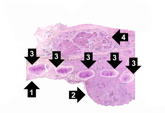
25 KB | This is a low-power photomicrograph of bronchus showing normal mucosa (1) with transition to carcinoma (2). Note the bronchial cartilage (3) and the invasion of tumor through the entire wall of the bronchus with tumor extending to the serosal surface (4). | 1 |
| 04:03, 21 August 2013 | IPLab10Candidiasis3.jpg (file) |  |
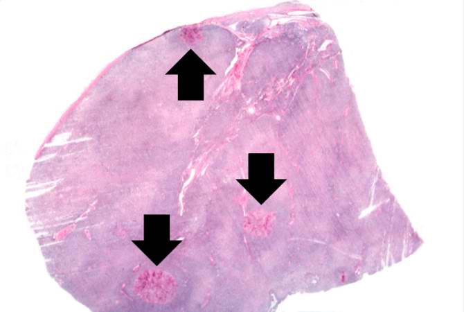
25 KB | This is a low-power photomicrograph of lymph node with three prominent areas of Candida colonies (arrows). Even at this low magnification, the purple-staining yeast and pseudohyphae can be easily seen. This section was stained with Periodic Acid-Schiff... | 1 |
| 04:15, 21 August 2013 | IPLab10Blasto3.jpg (file) |  |
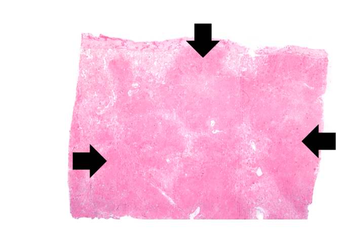
26 KB | This is a low-power photomicrograph of lung showing many areas of consolidation (arrows). | 1 |
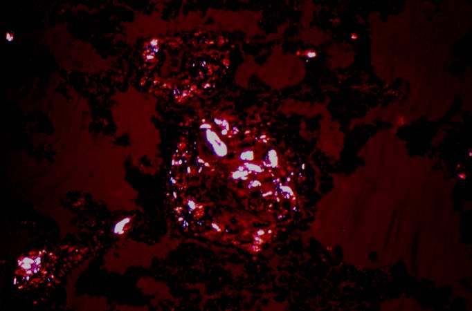
| 03:48, 19 August 2013 | IPLab3ForeignBodyGranuloma5.jpg (file) |  |
26 KB | This is a fully-polarized view of lung showing numerous birefringent particles. | 1 |