File list
This special page shows all uploaded files.
| Date | Name | Thumbnail | Size | Description | Versions |
|---|---|---|---|---|---|
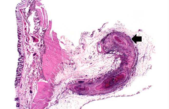
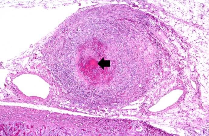
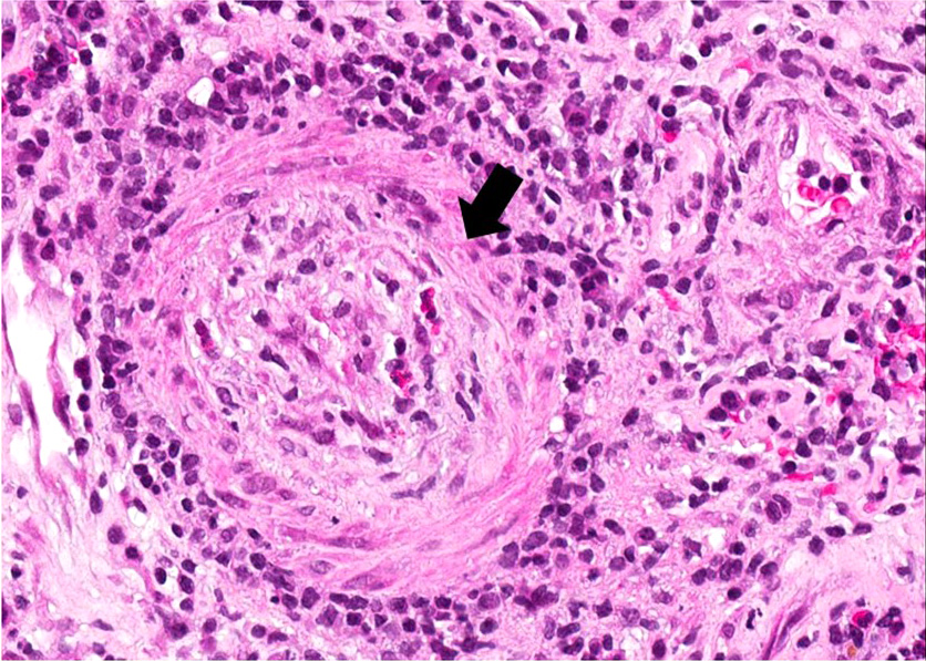
| 17:56, 20 August 2013 | IPLab6PAN4.jpg (file) |  |
51 KB | This is a low-power photomicrograph of a mesenteric vessel from this case of polyarteritis nodosa (arrow). The vessel is completely occluded by thrombotic material and the vessel wall is infiltrated with inflammatory cells. | 1 |
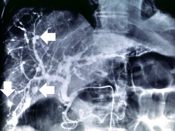
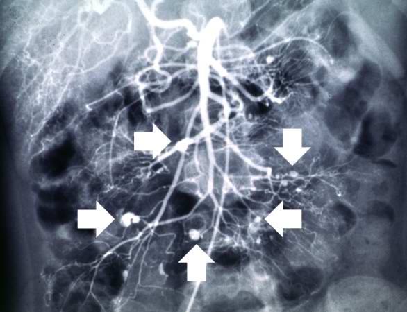
| 17:56, 20 August 2013 | IPLab6PAN3.jpg (file) |  |
36 KB | This angiogram of the kidneys demonstrates numerous aneurysmal dilatations in the renal circulation (arrows). | 1 |
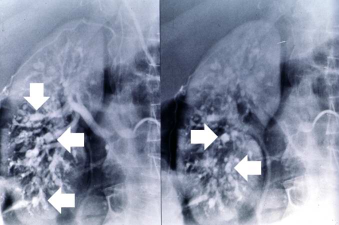
| 17:55, 20 August 2013 | IPLab6PAN2.jpg (file) |  |
36 KB | This angiogram of the liver also demonstrates numerous aneurysms throughout the hepatic circulation (arrows). | 1 |
| 18:00, 20 August 2013 | IPLab6PAN13.jpg (file) |  |
85 KB | This is a high-power photomicrograph of the affected vessel in the heart. The vessel lumen is completely occluded. | 1 |
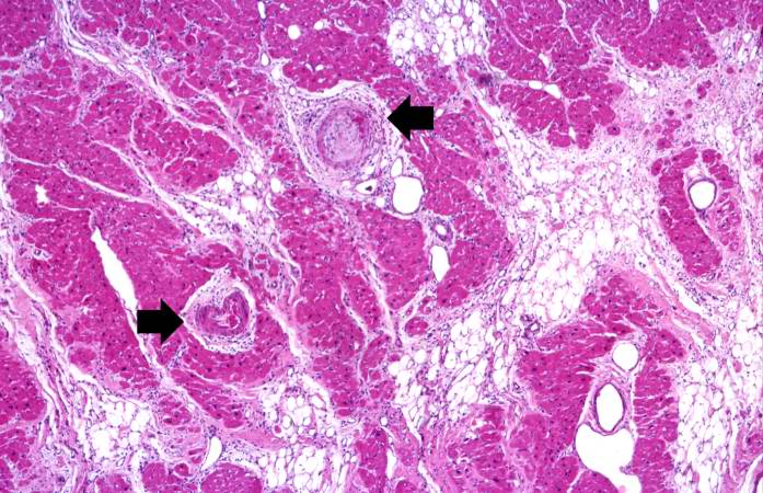
| 18:00, 20 August 2013 | IPLab6PAN12.jpg (file) |  |
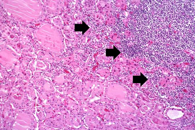
88 KB | This is a higher-power photomicrograph of the affected vessels in the heart (arrows). There are areas of fibrosis (old infarcts) in the myocardium adjacent to these affected vessels. | 1 |
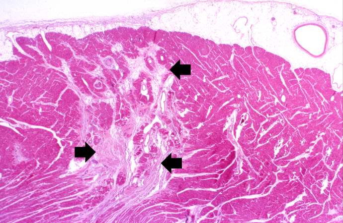
| 17:59, 20 August 2013 | IPLab6PAN11.jpg (file) |  |
69 KB | This is a low-power photomicrograph of the heart. There are areas of fibrosis in the myocardium (arrows). Note that the large epicardial coronary artery is normal. | 1 |
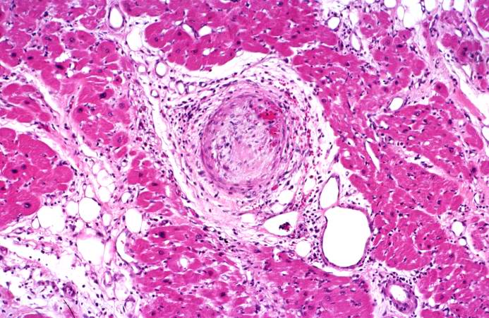
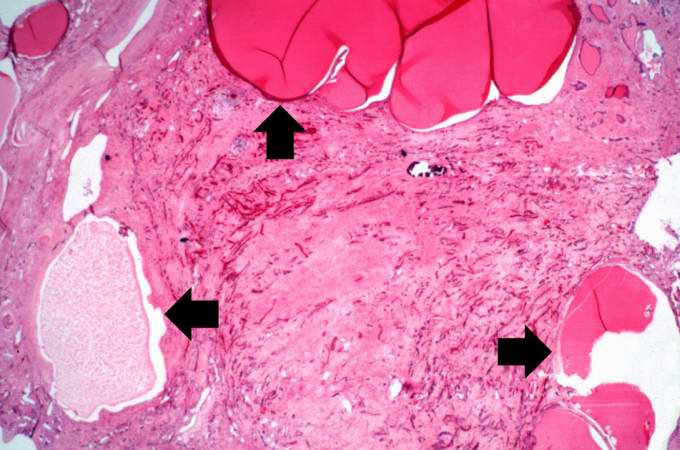
| 17:59, 20 August 2013 | IPLab6PAN10.jpg (file) |  |
76 KB | This is a higher-power photomicrograph of the affected vessel from the previous image. The vessel wall is infiltrated with inflammatory cells and the vessel lumen is completely occluded (arrow). | 1 |
| 17:55, 20 August 2013 | IPLab6PAN1.jpg (file) |  |
36 KB | This angiogram of the abdominal viscera demonstrates numerous aneurysms throughout the mesenteric circulation (arrows). | 1 |
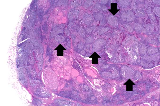
| 17:47, 20 August 2013 | IPLab6Hashimoto9.jpg (file) | 111 KB | This high-power photomicrograph shows more clearly the lymphocytes and plasma cells surrounding the thyroid gland epithelium. Large, eosinophilic, degenerating thyroid gland cells (Hurthle cells) can be seen in this section (arrows). | 1 | |
| 17:47, 20 August 2013 | IPLab6Hashimoto8.jpg (file) | 94 KB | This is a high-power photomicrograph showing the lymphocytes and plasma cells surrounding the thyroid gland epithelium. | 1 | |
| 17:46, 20 August 2013 | IPLab6Hashimoto7.jpg (file) | 87 KB | This is a high-power photomicrograph showing the inflammatory cells infiltrating into the residual thyroid tissue (arrows). | 1 | |
| 17:46, 20 August 2013 | IPLab6Hashimoto6.jpg (file) | 75 KB | This is another higher-power photomicrograph of thyroid from this case showing the inflammatory cells and the residual thyroid tissue. | 1 | |
| 23:29, 8 July 2020 | IPLab6Hashimoto5b.JPG (file) | 331 KB | 1 | ||
| 17:44, 20 August 2013 | IPLab6Hashimoto5.jpg (file) | 76 KB | This is a higher-power photomicrograph of thyroid from this case showing the inflammatory cells and the residual thyroid tissue. | 1 | |
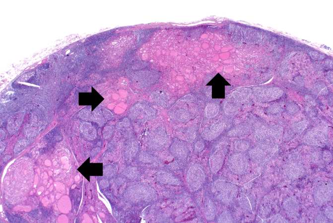
| 17:43, 20 August 2013 | IPLab6Hashimoto4.jpg (file) | 58 KB | This is another view of thyroid gland filled with inflammatory cells forming germinal centers (arrows). | 1 | |
| 17:43, 20 August 2013 | IPLab6Hashimoto3.jpg (file) | 55 KB | This is a higher-power photomicrograph of thyroid from this case. Note the large number of blue-staining inflammatory cells in this tissue. These cells appear to be forming germinal centers. Some residual thyroid gland tissue can be seen in this sectio... | 1 | |
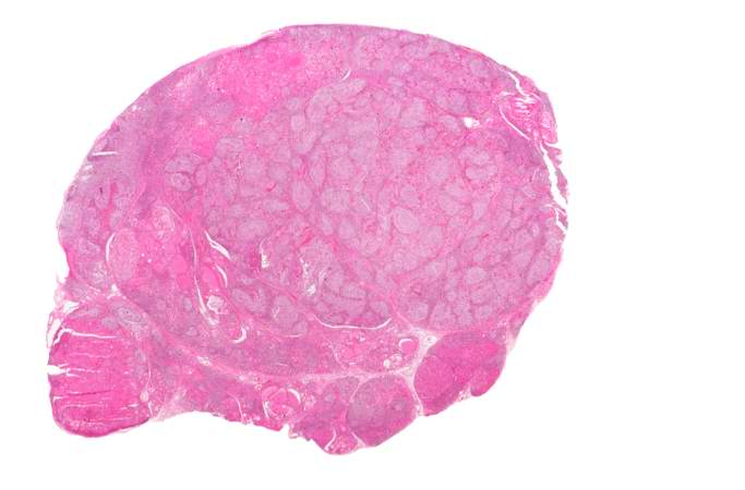
| 17:42, 20 August 2013 | IPLab6Hashimoto2.jpg (file) | 27 KB | This is a low-power photomicrograph of thyroid from this case. Note that the tissue is more cellular than one would expect and there does not appear to be normal colloid-filled blue spaces in this gland. | 1 | |
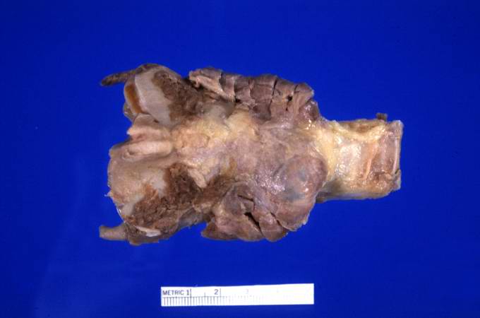
| 17:42, 20 August 2013 | IPLab6Hashimoto1.jpg (file) | 20 KB | This is a gross photograph of thyroid gland taken at autopsy. The gland is only slightly enlarged and has a firm texture. | 1 | |
| 21:38, 8 July 2020 | IPLab6GravesDisease7b.jpg (file) |  |
338 KB | 1 | |
| 23:13, 8 July 2020 | IPLab6GravesDisease5x.jpg (file) |  |
278 KB | 1 | |
| 23:13, 8 July 2020 | IPLab6GravesDisease3x.jpg (file) |  |
277 KB | 1 | |
| 21:35, 8 July 2020 | IPLab6GravesDisease1b.jpg (file) |  |
393 KB | 1 | |
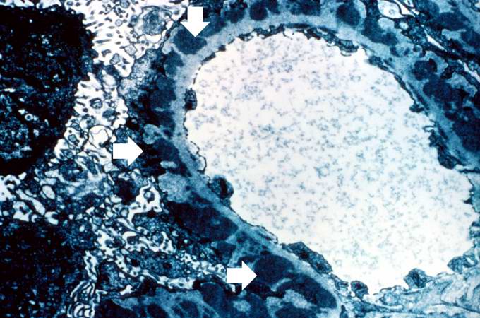
| 20:30, 20 August 2013 | IPLab6GN9.jpg (file) |  |
74 KB | This electron micrograph demonstrates scattered subepithelial dense deposits (arrows) and a polymorphonuclear leukocyte in the lumen. | 1 |
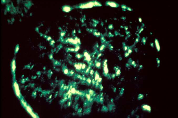
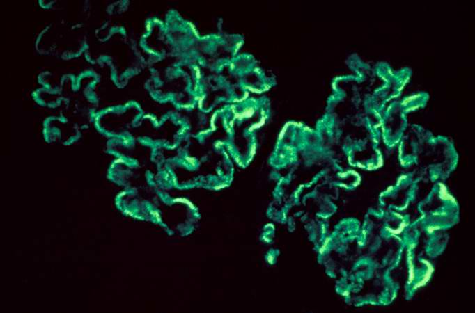
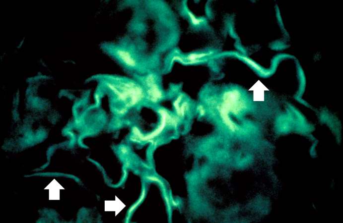
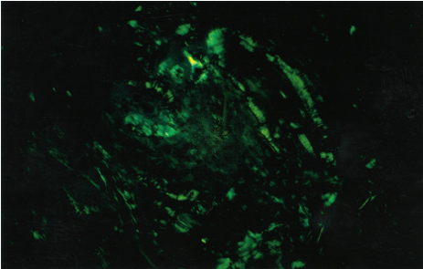
| 20:30, 20 August 2013 | IPLab6GN8.jpg (file) |  |
31 KB | This immunofluorescent photomicrograph of a glomerulus from a case of acute poststreptococcal glomerulonephritis shows a granular immunofluorescence pattern consistent with immune complex disease. The primary antibody used for this staining was specifi... | 1 |
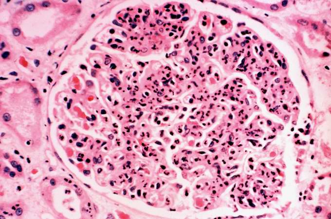
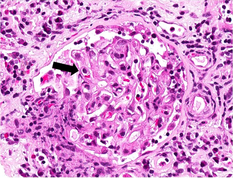
| 20:29, 20 August 2013 | IPLab6GN7.jpg (file) |  |
62 KB | This is a photomicrograph of a glomerulus from another case with acute poststreptococcal glomerulonephritis. In this case the immune complex glomerular disease is ongoing with necrosis and accumulation of neutrophils in the glomerulus. | 1 |
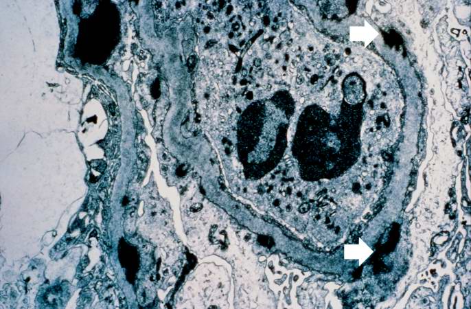
| 20:29, 20 August 2013 | IPLab6GN6.jpg (file) |  |
66 KB | This is an electron micrograph of subepithelial granular electron dense deposits (arrows) which correspond to the granular immunofluorescence seen in the previous image. | 1 |
| 20:28, 20 August 2013 | IPLab6GN5.jpg (file) |  |
30 KB | This is an immunofluorescent photomicrograph of granular membranous immunofluorescence (immune complex disease). The antibody used for these studies was specific for IgG. | 1 |
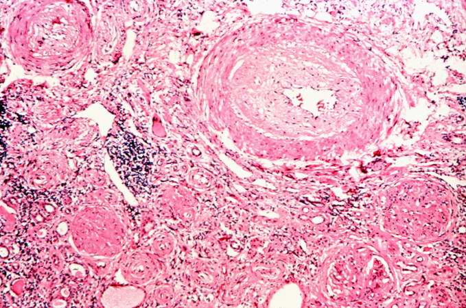
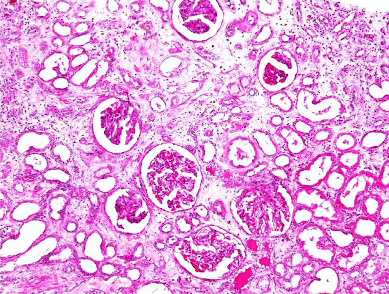
| 20:28, 20 August 2013 | IPLab6GN4.jpg (file) |  |
87 KB | This is a photomicrograph of interstitial and vascular lesions in end stage renal disease. | 1 |
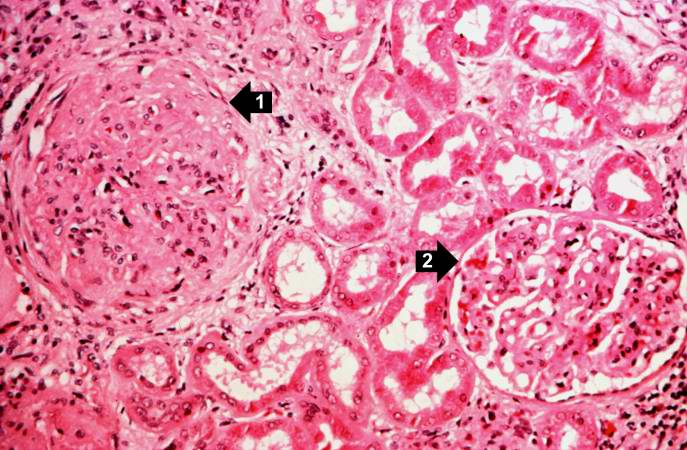
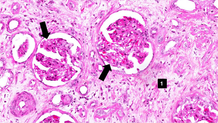
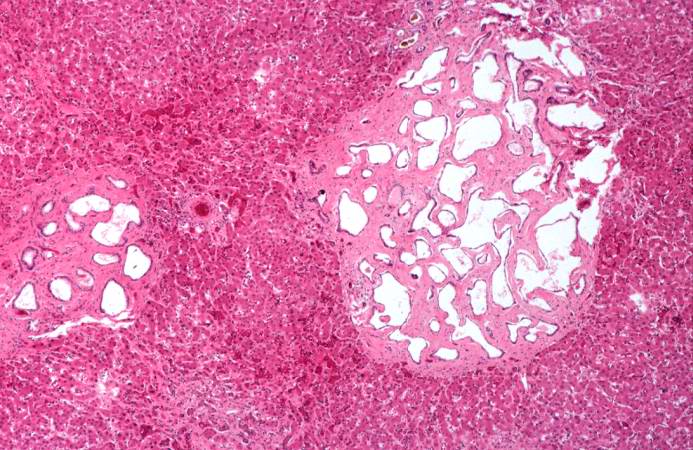
| 20:27, 20 August 2013 | IPLab6GN3.jpg (file) |  |
71 KB | This is a higher-power photomicrograph of hyalinized glomeruli (1) and glomeruli with thickened basement membranes (2). | 1 |
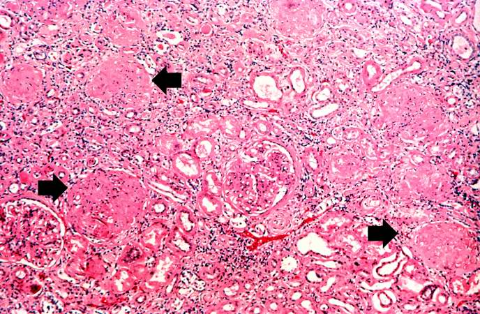
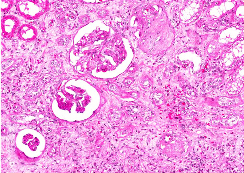
| 20:27, 20 August 2013 | IPLab6GN2.jpg (file) |  |
103 KB | This is a higher-power photomicrograph of hyalinized glomeruli (arrows) and glomeruli with thick basement membranes. | 1 |
| 20:30, 20 August 2013 | IPLab6GN10.jpg (file) |  |
29 KB | For comparison this is an immunofluorescent photomicrograph of a glomerulus from a patient with Goodpasture's syndrome. The linear (arrows) immunofluorescence is characteristic of Goodpasture's syndrome. | 1 |
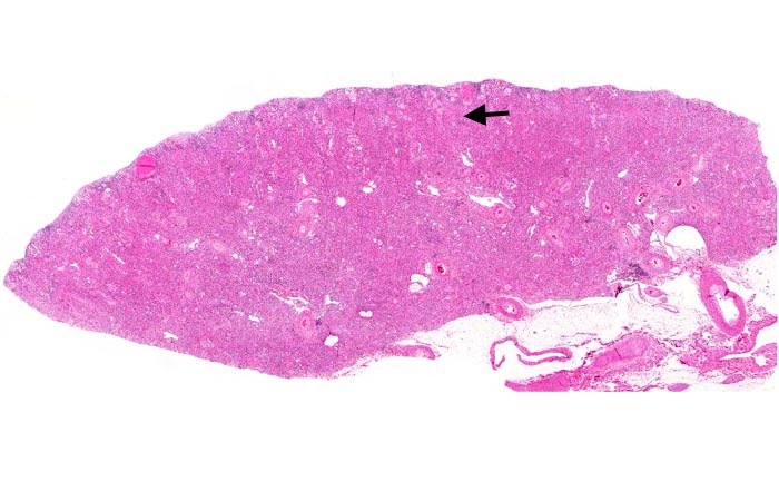
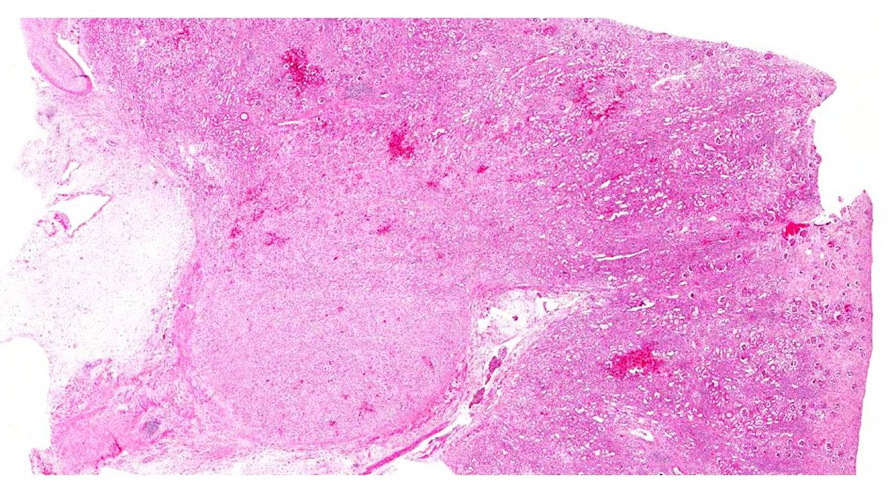
| 20:43, 20 August 2013 | IPLab6GN1.jpg (file) |  |
72 KB | This is a low-power photomicrograph of a saggital section of end stage chronic glomerulonephritis (GN). Note the marked thinning of the cortex (arrow). | 1 |
| 00:00, 9 July 2020 | IPLab6ChronicRejection9b.jpg (file) |  |
315 KB | 1 | |
| 00:00, 9 July 2020 | IPLab6ChronicRejection5b.jpg (file) |  |
336 KB | 1 | |
| 00:00, 9 July 2020 | IPLab6ChronicRejection4b.jpg (file) |  |
352 KB | 1 | |
| 00:00, 9 July 2020 | IPLab6ChronicRejection3b.jpg (file) |  |
390 KB | 1 | |
| 23:59, 8 July 2020 | IPLab6ChronicRejection2b.jpg (file) |  |
406 KB | 1 | |
| 23:59, 8 July 2020 | IPLab6ChronicRejection1b.jpg (file) |  |
267 KB | 1 | |
| 00:19, 9 July 2020 | IPLab6Amyloid8b.jpg (file) |  |
109 KB | 1 | |
| 17:53, 19 August 2013 | IPLab5PolycysticKidney9.jpg (file) |  |
81 KB | This photomicrograph of liver demonstrates the histologic appearance of these cysts. | 1 |
| 19:35, 8 July 2020 | IPLab5PolycysticKidney8b.jpg (file) |  |
439 KB | 1 | |
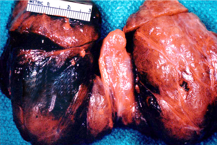
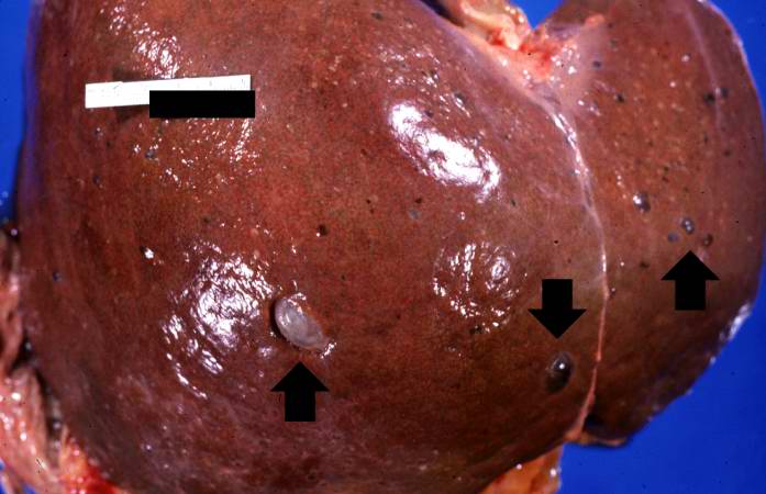
| 17:53, 19 August 2013 | IPLab5PolycysticKidney8.jpg (file) |  |
42 KB | This is a gross photograph of the liver from this patient. Multiple cysts can be seen on the surface of this liver (arrows). | 1 |
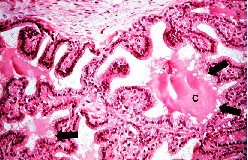
| 17:52, 19 August 2013 | IPLab5PolycysticKidney7.jpg (file) |  |
66 KB | This high-power photomicrograph shows abnormal glomeruli (arrows) and some tubules. | 1 |
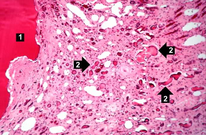
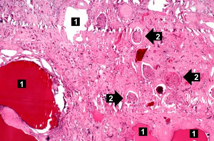
| 17:52, 19 August 2013 | IPLab5PolycysticKidney6.jpg (file) |  |
66 KB | This is a higher-power photomicrograph of polycystic kidney showing the edge of a large cyst (1). In this section there are numerous tubules and dilated collecting ducts (2) that are filled with the same red proteinaceous material as the larger cysts. | 1 |
| 17:51, 19 August 2013 | IPLab5PolycysticKidney5.jpg (file) |  |
53 KB | This is another low-power photomicrograph of an H&E-stained section from this polycystic kidney. Again note the large cystic structures (arrows)and the fibrous connective tissue throughout this section. | 1 |
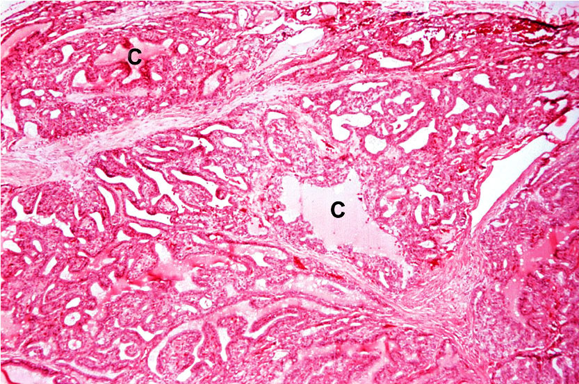
| 17:51, 19 August 2013 | IPLab5PolycysticKidney4.jpg (file) |  |
69 KB | This is a low-power photomicrograph of an H&E-stained section from this polycystic kidney. Note the large cystic structures (1), the few residual glomeruli (2), and the fibrous connective tissue throughout this section. | 1 |
| 19:33, 8 July 2020 | IPLab5PolycysticKidney3b.jpg (file) |  |
263 KB | 1 | |
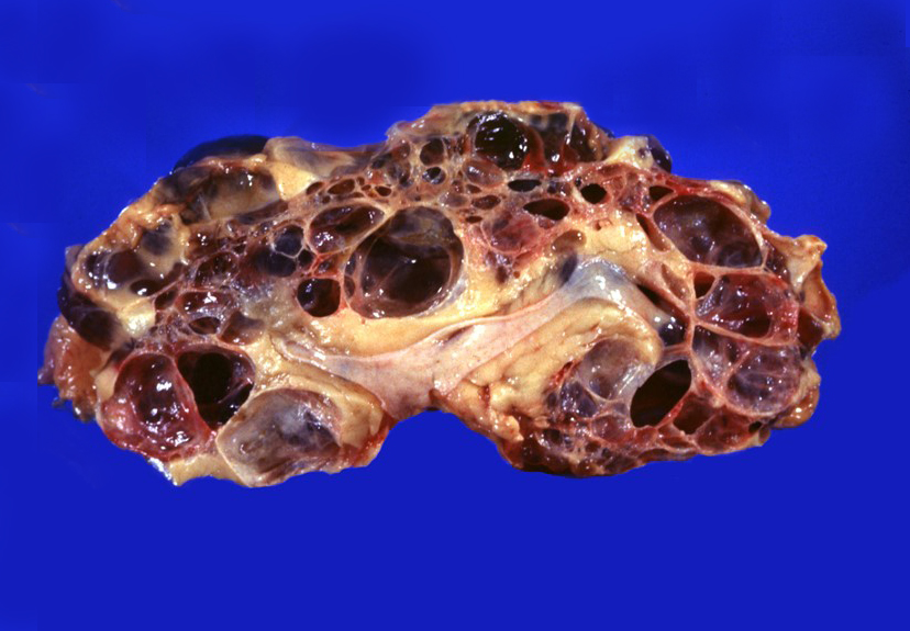
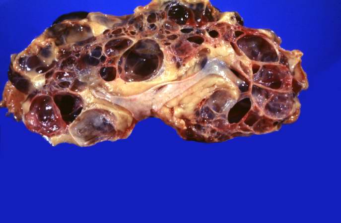
| 17:49, 19 August 2013 | IPLab5PolycysticKidney3.jpg (file) |  |
33 KB | This is a gross photograph of a cut section from one of these polycystic kidneys. Note that the renal parenchyma is almost completely replaced by cystic structures. | 1 |
| 19:32, 8 July 2020 | IPLab5PolycysticKidney2b.jpg (file) |  |
213 KB | 1 | |
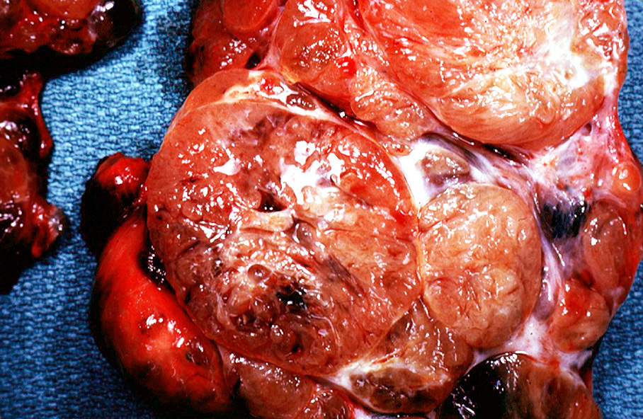
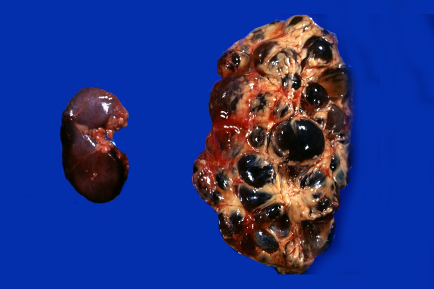
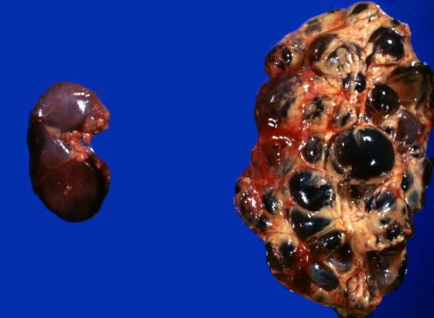
| 17:40, 19 August 2013 | IPLab5PolycysticKidney2.jpg (file) |  |
26 KB | This is a gross photograph of the kidneys from this case. Note that both kidneys contain multiple large cysts (arrows). | 1 |