File list
This special page shows all uploaded files.
| Date | Name | Thumbnail | Size | Description | Versions |
|---|---|---|---|---|---|
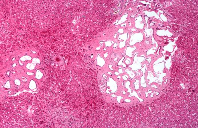
| 17:54, 19 August 2013 | IPLab5PolycysticKidney11.jpg (file) |  |
68 KB | This is a higher-power photomicrograph of liver cyst. These cystic structures are lined by biliary epithelium. | 1 |
| 20:11, 20 August 2013 | IPLab6TB4.jpg (file) |  |
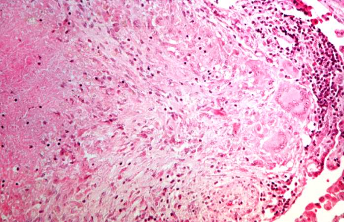
68 KB | This is a higher-power photomicrograph of a TB granuloma. The area of caseous necrosis is on the left side of the image, there are multinucleated giant cells and epithelioid macrophages throughout the remainder of the tissue. | 1 |
| 16:03, 19 August 2013 | IPLab2Atrophy4.jpg (file) |  |
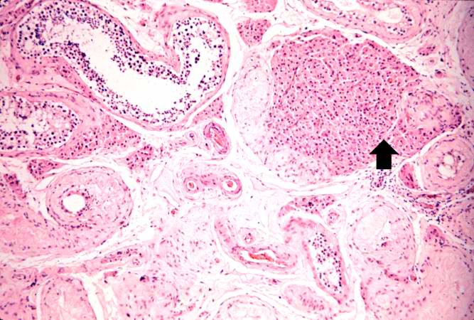
68 KB | This is a higher-power photomicrograph indicating loss of testicular parenchymal tissue. There are very few recognizable spermatic cells in this tissue. The cluster of cells in the upper right is a focus of interstitial or Leydig cells (arrow). These c... | 1 |
| 18:28, 19 August 2013 | IPLab5Antitrypsin12.jpg (file) |  |
68 KB | This is a high-power photomicrograph of liver stained with periodic-acid Schiff's (PAS) stain. This demonstrates the PAS-positive granules of defective alpha 1-antitrypsin that accumulate in the Golgi of hepatocytes (arrows). | 1 |
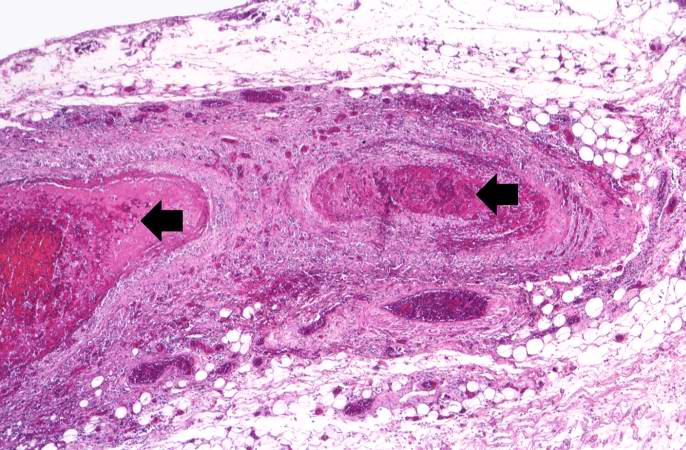
| 17:59, 20 August 2013 | IPLab6PAN11.jpg (file) |  |
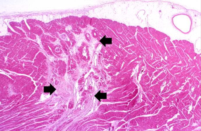
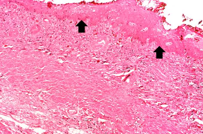
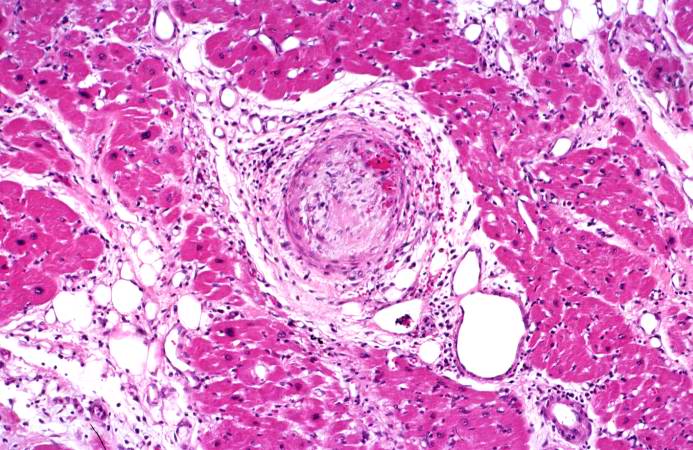
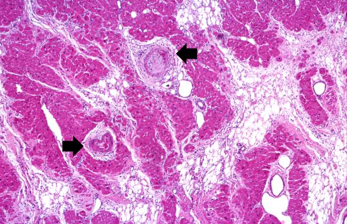
69 KB | This is a low-power photomicrograph of the heart. There are areas of fibrosis in the myocardium (arrows). Note that the large epicardial coronary artery is normal. | 1 |
| 18:07, 19 August 2013 | IPLab5Antitrypsin4.jpg (file) |  |
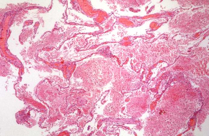
69 KB | This is a gross photograph of the bronchi and lungs. Note the hemorrhage in the bronchi and in the lung parenchyma. | 1 |
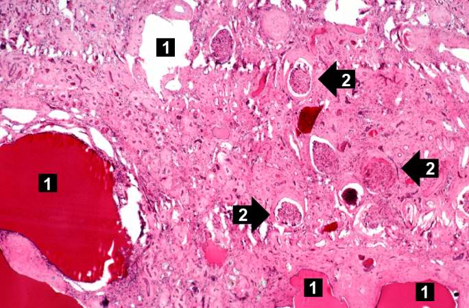
| 17:51, 19 August 2013 | IPLab5PolycysticKidney4.jpg (file) |  |
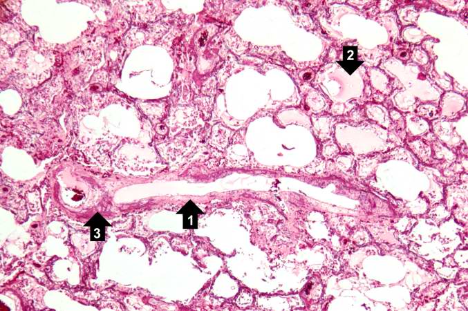
69 KB | This is a low-power photomicrograph of an H&E-stained section from this polycystic kidney. Note the large cystic structures (1), the few residual glomeruli (2), and the fibrous connective tissue throughout this section. | 1 |
| 15:28, 19 August 2013 | IPLab2Hyperplasia4.jpg (file) |  |
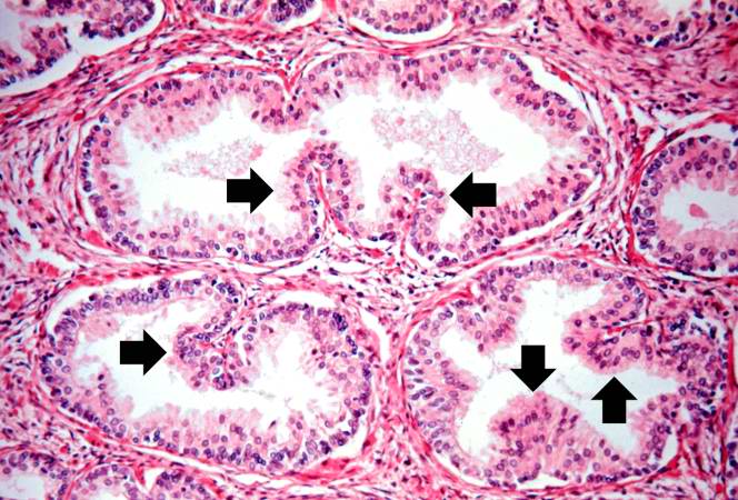
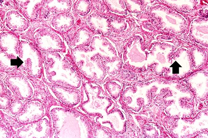
69 KB | The dilated glands (arrows) make up the major portion of the prostate tissue and there is compression of the stroma. | 1 |
| 18:27, 19 August 2013 | IPLab5Antitrypsin9.jpg (file) |  |
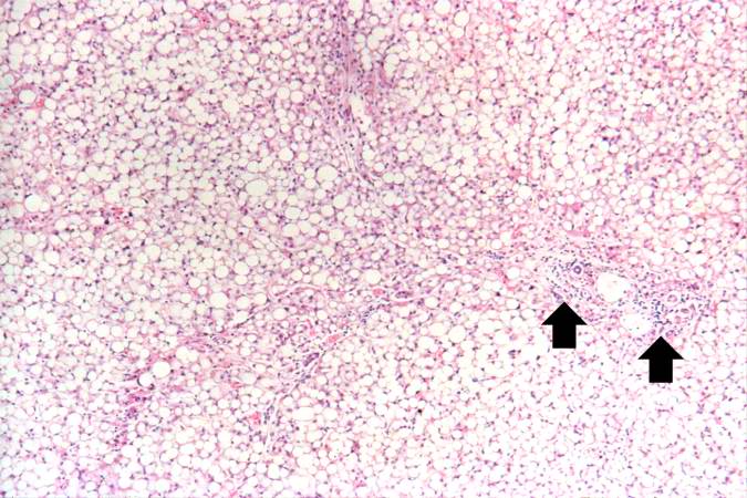
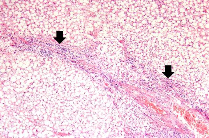
70 KB | This is a low-power photomicrograph of an H&E-stained section of liver. There are increased numbers of inflammatory cells in the periportal region (arrow) and the central vein areas are pale. | 1 |
| 20:27, 20 August 2013 | IPLab6GN3.jpg (file) |  |
71 KB | This is a higher-power photomicrograph of hyalinized glomeruli (1) and glomeruli with thickened basement membranes (2). | 1 |
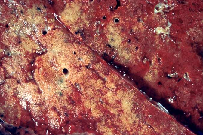
| 16:32, 19 August 2013 | IPLab2Calcification1.jpg (file) |  |
72 KB | This is a gross photograph of the cut section of the patient's lung showing evidence of severe metastatic calcification. The lung tissue has a rough, firm appearance with open airways. | 1 |
| 15:08, 20 August 2013 | IPLab5Gaucher2.jpg (file) |  |
72 KB | This is a cut section of spleen from this case. Again note the fine granular appearance to the tissue. | 1 |
| 20:43, 20 August 2013 | IPLab6GN1.jpg (file) |  |
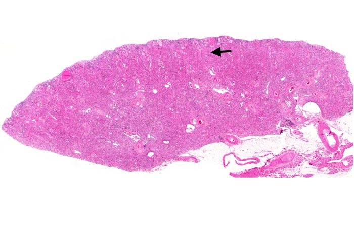
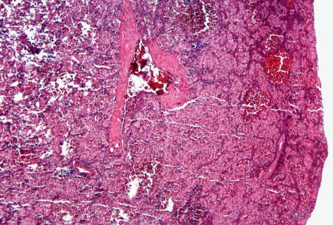
72 KB | This is a low-power photomicrograph of a saggital section of end stage chronic glomerulonephritis (GN). Note the marked thinning of the cortex (arrow). | 1 |
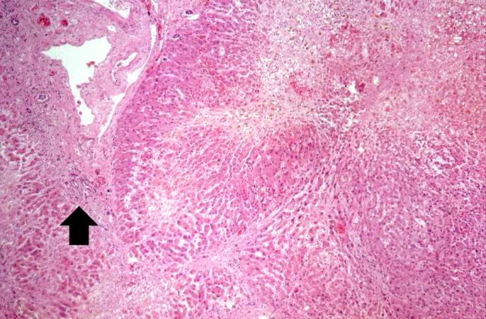
| 20:10, 20 August 2013 | IPLab6TB3.jpg (file) |  |
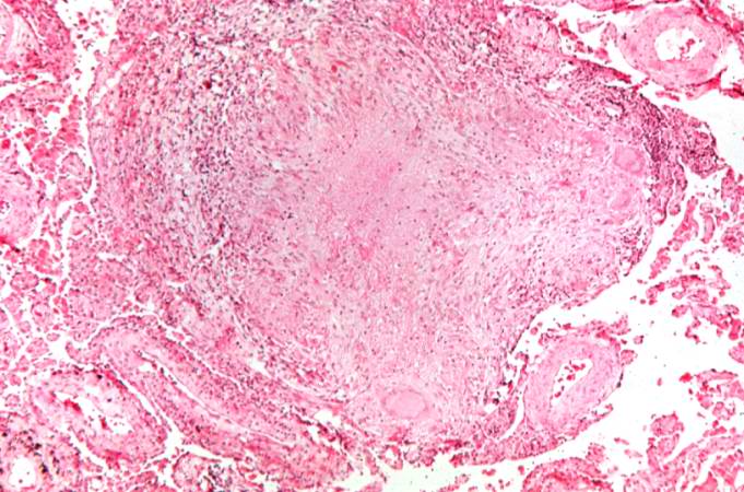
72 KB | This is a higher-power photomicrograph of a TB granuloma. Note the eosinophilic material in the center of this granuloma (caseous necrosis) and the epithelioid macrophages and giant cells around the periphery. | 1 |
| 16:55, 19 August 2013 | IPLab2FattyChange3.jpg (file) |  |
73 KB | Another low-power photomicrograph illustrates again the pale, washed-out appearance of this tissue. Notice the numerous holes throughout the tissue. There are accumulations of inflammatory cells (arrows) around portal tracts. | 1 |
| 15:27, 20 August 2013 | IPLab5DM4.jpg (file) |  |
74 KB | This is a high-power photomicrograph of two glomeruli with intercapillary glomerulosclerosis (arrows). | 1 |
| 20:30, 20 August 2013 | IPLab6GN9.jpg (file) |  |
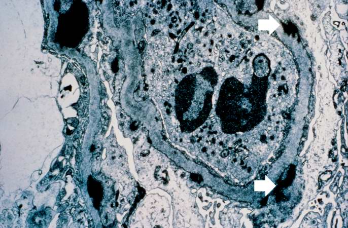
74 KB | This electron micrograph demonstrates scattered subepithelial dense deposits (arrows) and a polymorphonuclear leukocyte in the lumen. | 1 |
| 15:29, 19 August 2013 | IPLab2Hyperplasia7.jpg (file) |  |
74 KB | A higher-power view shows the papillary folds (arrows) produced by the hyperplastic epithelium projecting into the lumen of the gland. While these papillary folds project into the lumen of the gland, there is no extension through the glandular basement... | 1 |
| 17:21, 19 August 2013 | IPLab5Neurofibromatosis7.jpg (file) |  |
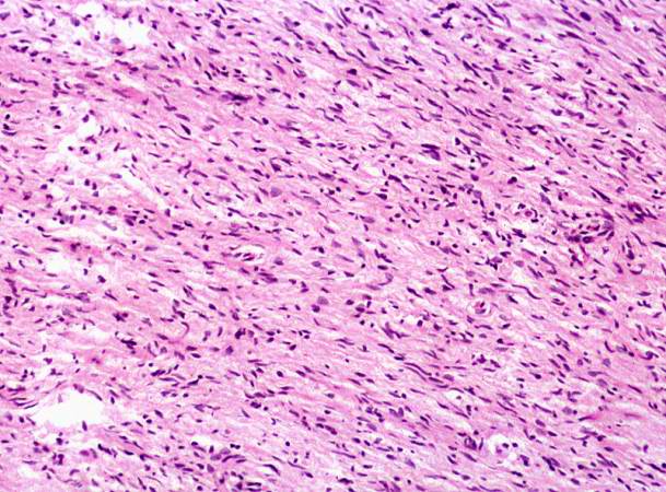
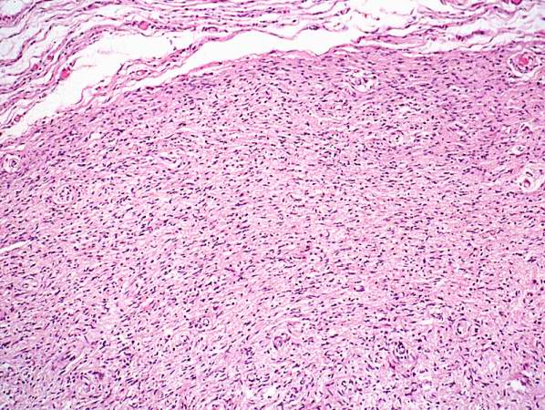
74 KB | This higher-power photomicrograph of the neurofibroma shows more clearly the elongated cells (primarily Schwann cells) that make up this tumor. | 1 |
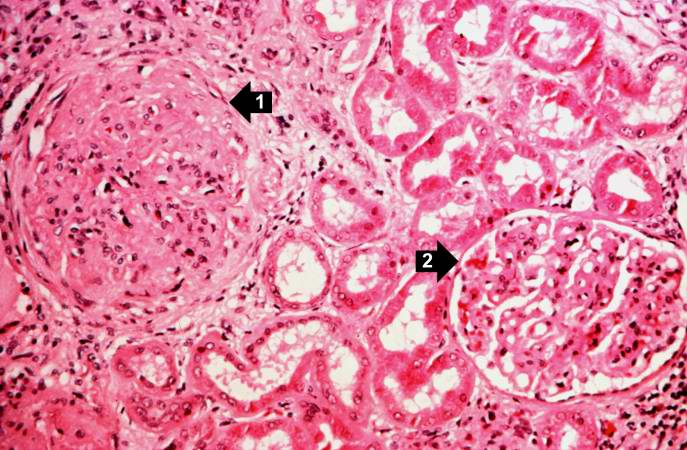
| 15:27, 20 August 2013 | IPLab5DM5.jpg (file) |  |
74 KB | This is a photomicrograph of a glomerulus with nodular glomerulosclerosis (1). Also note the intertubular fibrosis and the changes in the blood vessels (2). | 1 |
| 16:56, 19 August 2013 | IPLab2FattyChange5.jpg (file) |  |
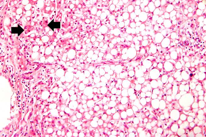
75 KB | This higher-power photomicrograph of the centrilobular area gives the appearance of fatty tissue, as indicated by many empty spaces. Very few normal liver cells can be seen in this slide. A few more normal-appearing hepatocytes are present at the left ... | 1 |
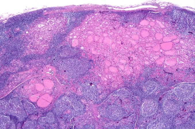
| 17:46, 20 August 2013 | IPLab6Hashimoto6.jpg (file) | 75 KB | This is another higher-power photomicrograph of thyroid from this case showing the inflammatory cells and the residual thyroid tissue. | 1 | |
| 17:44, 20 August 2013 | IPLab6Hashimoto5.jpg (file) | 76 KB | This is a higher-power photomicrograph of thyroid from this case showing the inflammatory cells and the residual thyroid tissue. | 1 | |
| 15:44, 19 August 2013 | IPLab2Metaplasia5.jpg (file) |  |
76 KB | In areas adjacent to the normal transitional epithelium, there are areas of epithelium (arrows) where the epithelial cells have the character of normal squamous epithelium as found in the dermis. However, squamous epithelium is not normal in the renal ... | 1 |
| 17:59, 20 August 2013 | IPLab6PAN10.jpg (file) |  |
76 KB | This is a higher-power photomicrograph of the affected vessel from the previous image. The vessel wall is infiltrated with inflammatory cells and the vessel lumen is completely occluded (arrow). | 1 |
| 16:56, 19 August 2013 | IPLab2FattyChange4.jpg (file) |  |
77 KB | A higher-power photomicrograph illustrates more clearly the inflammatory cells (arrows) around the portal areas. | 1 |
| 18:28, 19 August 2013 | IPLab5Antitrypsin11.jpg (file) |  |
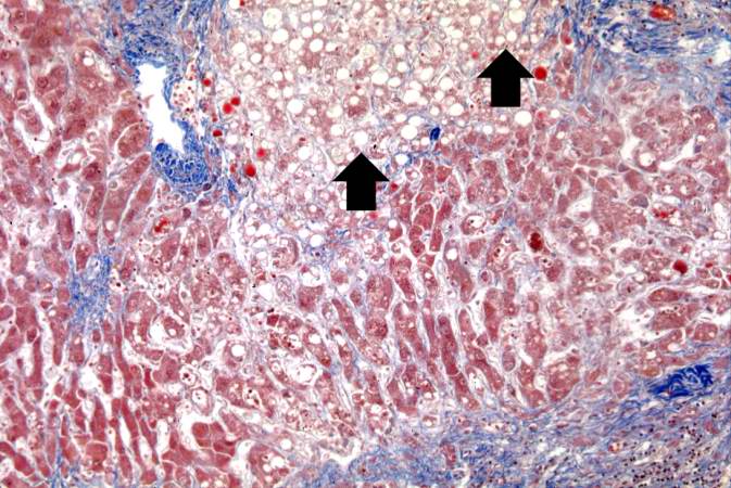
77 KB | This is a higher-power photomicrograph of a trichrome-stained section of liver. This section demonstrates the fibrosis (blue material) and the fatty change (arrows). | 1 |
| 16:58, 19 August 2013 | IPLab2FattyChange8.jpg (file) |  |
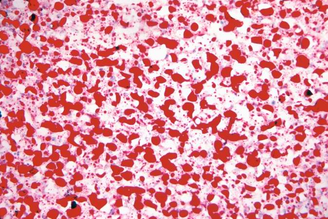
78 KB | An oil red O stain for fat was performed on a frozen section of this liver tissue. The red droplets represent fat in the tissue which is typical of fatty degeneration in the liver. By using frozen sections the tissues do not have to be dehydrated throu... | 1 |
| 14:55, 20 August 2013 | IPLab5Hemochromatosis8.jpg (file) |  |
78 KB | This higher-power view of liver stained with Prussian blue demonstrates the marked accumulation of iron within the parenchymal cells (1) and in the Kupffer cells in the periportal area (2). | 1 |
| 16:33, 19 August 2013 | IPLab2Calcification3.jpg (file) |  |
78 KB | A higher-power photomicrograph shows a blood vessel cut in longitudinal section (1). Several of the alveoli are filled with a pink-staining proteinaceous fluid (2) indicative of pulmonary edema. The alveolar septa and the wall of the blood vessel have ... | 1 |
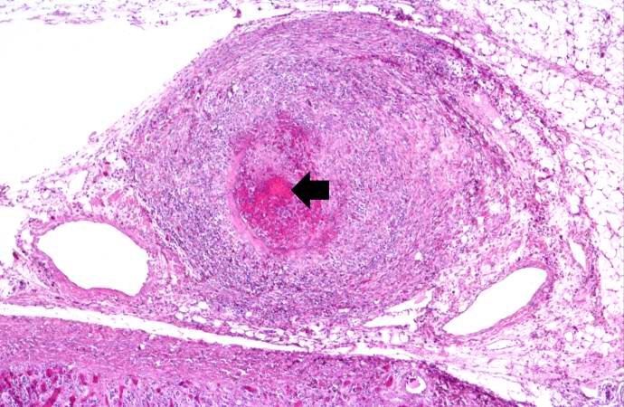
| 17:56, 20 August 2013 | IPLab6PAN5.jpg (file) |  |
79 KB | This is a higher-power photomicrograph of this mesenteric vessel. Note the thrombotic material occluding the vessel (arrows) and the inflammatory cell infiltrate in the wall of the vessel and in the surrounding adventitia. | 1 |
| 18:27, 19 August 2013 | IPLab5Antitrypsin10.jpg (file) |  |
80 KB | This is a low-power photomicrograph of a trichrome-stained section of liver. There is bridging fibrosis (blue material) between portal regions. | 1 |
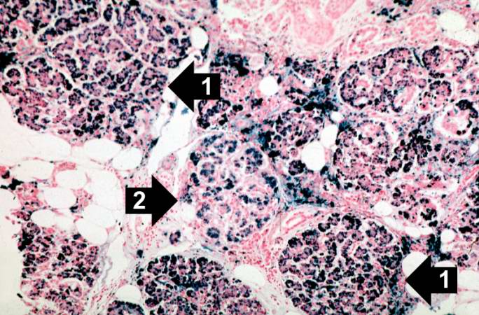
| 14:57, 20 August 2013 | IPLab5Hemochromatosis11.jpg (file) |  |
81 KB | This is a histologic section of pancreas from this case stained for iron (Prussian blue). Note the accumulation of iron in the parenchymal cells (1). There is also iron in the pancreatic islets (2). | 1 |
| 17:53, 19 August 2013 | IPLab5PolycysticKidney9.jpg (file) |  |
81 KB | This photomicrograph of liver demonstrates the histologic appearance of these cysts. | 1 |
| 18:00, 20 August 2013 | IPLab6PAN13.jpg (file) |  |
85 KB | This is a high-power photomicrograph of the affected vessel in the heart. The vessel lumen is completely occluded. | 1 |
| 17:58, 20 August 2013 | IPLab6PAN8.jpg (file) |  |
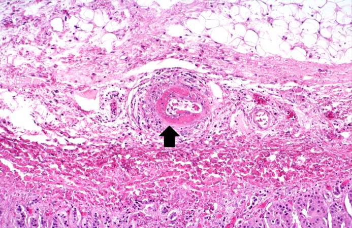
86 KB | This is a high-power photomicrograph of a small vessel with a rim of fibrinoid necrosis (arrow). | 1 |
| 15:28, 19 August 2013 | IPLab2Hyperplasia5.jpg (file) |  |
86 KB | Note these glands, which exhibit hyperplasia of the glandular epithelium. The infolding of the glandular epithelial cells forms papillary projections (arrows) into the lumen of the gland. | 1 |
| 15:11, 20 August 2013 | IPLab5Gaucher4.jpg (file) |  |
86 KB | This is a photomicrograph of the spleen from this case. There is very little if any white pulp evident in this section. | 1 |
| 15:11, 20 August 2013 | IPLab5Gaucher6.jpg (file) |  |
87 KB | This is another high-power photomicrograph of the spleen from this case. At this power it is easier to see the large eosinophilic cells. | 1 |
| 20:28, 20 August 2013 | IPLab6GN4.jpg (file) |  |
87 KB | This is a photomicrograph of interstitial and vascular lesions in end stage renal disease. | 1 |
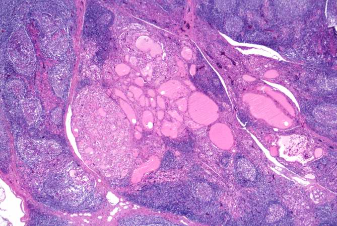
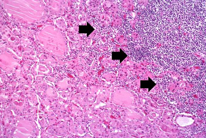
| 17:46, 20 August 2013 | IPLab6Hashimoto7.jpg (file) | 87 KB | This is a high-power photomicrograph showing the inflammatory cells infiltrating into the residual thyroid tissue (arrows). | 1 | |
| 18:00, 20 August 2013 | IPLab6PAN12.jpg (file) |  |
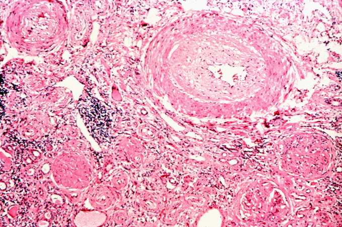
88 KB | This is a higher-power photomicrograph of the affected vessels in the heart (arrows). There are areas of fibrosis (old infarcts) in the myocardium adjacent to these affected vessels. | 1 |
| 17:21, 19 August 2013 | IPLab5Neurofibromatosis6.jpg (file) |  |
89 KB | This is a higher-power photomicrograph of the neurofibroma demonstrating the loose pattern of elongated cells making up the tumor mass. | 1 |
| 15:11, 20 August 2013 | IPLab5Gaucher5.jpg (file) |  |
90 KB | This is a higher-power photomicrograph of the spleen from this case. Again there is no white pulp and the red pulp is filled with large eosinophilic cells. | 1 |
| 14:56, 20 August 2013 | IPLab5Hemochromatosis10.jpg (file) |  |
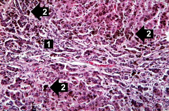
91 KB | This is a histologic section of pancreas from this case. It is difficult to appreciate at this magnification, but there is brown pigment in the pancreatic acinar cells. Note the islets of Langerhans (1). | 1 |
| 14:54, 20 August 2013 | IPLab5Hemochromatosis5.jpg (file) |  |
92 KB | This higher-power photomicrograph demonstrates the increased fibrosis in the periportal area (1) and the pigment accumulation (2). | 1 |
| 15:27, 20 August 2013 | IPLab5DM3.jpg (file) |  |
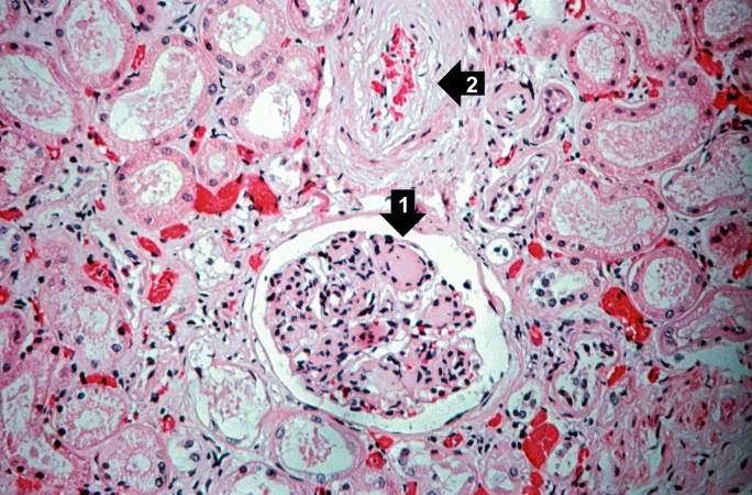
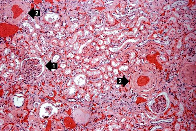
94 KB | This is a higher-power photomicrograph of the cortical region. In this region there is ischemic obsolescence of glomeruli and one glomerulus with nodular glomerulosclerosis (1). Also note the thickened walls of the blood vessels (2). | 1 |
| 14:53, 20 August 2013 | IPLab5Hemochromatosis4.jpg (file) |  |
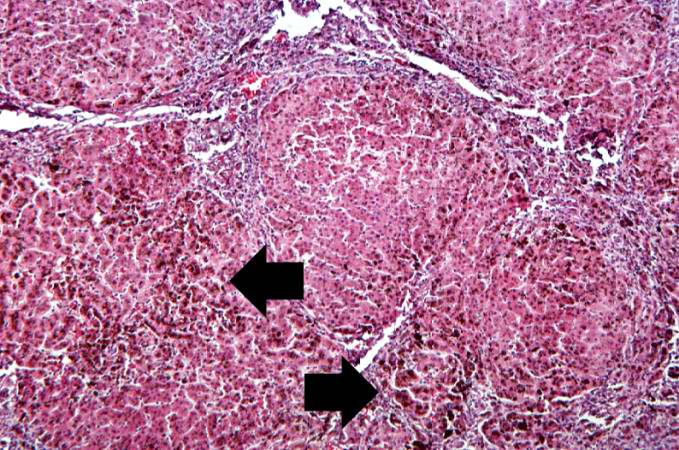
94 KB | This higher-power view of liver from this case demonstrates the nodules and the brown/black pigment within liver parenchymal cells (arrows). | 1 |
| 17:47, 20 August 2013 | IPLab6Hashimoto8.jpg (file) | 94 KB | This is a high-power photomicrograph showing the lymphocytes and plasma cells surrounding the thyroid gland epithelium. | 1 | |
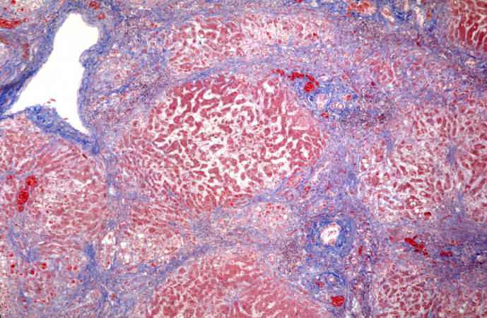
| 14:54, 20 August 2013 | IPLab5Hemochromatosis6.jpg (file) |  |
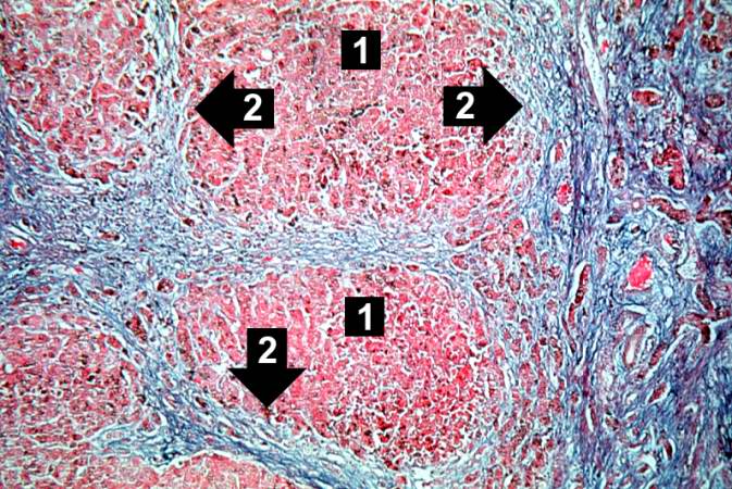
99 KB | This trichrome stain of liver section demonstrates the increased fibrous connective tissue in this liver. Note that the liver nodules (1) are surrounded by fibrous connective tissue (2). | 1 |